

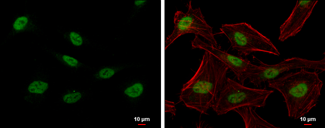
PTBP2 antibody detects polypyrimidine tract binding protein 2 protein at nucleus by immunofluorescent analysis. Sample: HeLa cells were fixed in 4% paraformaldehyde at RT for 15 min. Green: polypyrimidine tract binding protein 2 protein stained by PTBP2 antibody (GTX116704) diluted at 1:500. Red: phalloidin, a cytoskeleton marker, diluted at 1:200. Blue: Hoechst 33342 staining. Scale bar = 10 μm.
![PTBP2 antibody detects PTBP2 protein by immunofluorescent analysis. Sample: DIV9 rat E18 primary hippocampal neuron cells were fixed in 4% paraformaldehyde at RT for 15 min. Green: PTBP2 stained by PTBP2 antibody (GTX116704) diluted at 1:500. Red: beta Tubulin 3/ Tuj1, stained by beta Tubulin 3/ Tuj1 antibody [GT11710] (GTX631836) diluted at 1:500. Blue: Fluoroshield with DAPI (GTX30920).](https://www.genetex.com/upload/website/prouct_img/normal/GTX116704/GTX116704_41143_20181004_ICC_IF_R_w_23060519_964.webp "PTBP2 antibody detects PTBP2 protein by immunofluorescent analysis. Sample: DIV9 rat E18 primary hippocampal neuron cells were fixed in 4% paraformaldehyde at RT for 15 min. Green: PTBP2 stained by PTBP2 antibody (GTX116704) diluted at 1:500. Red: beta Tubulin 3/ Tuj1, stained by beta Tubulin 3/ Tuj1 antibody [GT11710] (GTX631836) diluted at 1:500. Blue: Fluoroshield with DAPI (GTX30920).")
 dilution: 1:2000")
 dilution: 1:500.
Antigen Retrieval: Trilogy? (EDTA based, pH 8.0) buffer, 15min")
 dilution: 1:500.
Antigen Retrieval: Trilogy? (EDTA based, pH 8.0) buffer, 15min")
 dilution: 1:500.
Antigen Retrieval: Trilogy? (EDTA based, pH 8.0) buffer, 15min")
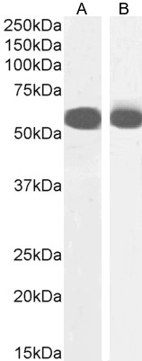
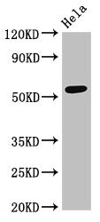
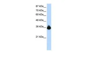
 was separated by 10% SDS-PAGE, and the membrane was blotted with PTBP2 antibody (GTX116704) diluted at 1:1000.")
PTBP2 antibody detects polypyrimidine tract binding protein 2 protein at nucleus by immunofluorescent analysis. Sample: HeLa cells were fixed in 4% paraformaldehyde at RT for 15 min. Green: polypyrimidine tract binding protein 2 protein stained by PTBP2 antibody (GTX116704) diluted at 1:500. Red: phalloidin, a cytoskeleton marker, diluted at 1:200. Blue: Hoechst 33342 staining. Scale bar = 10 μm.
PTBP2 antibody
GTX116704
ApplicationsImmunoFluorescence, Western Blot, ImmunoCytoChemistry, ImmunoHistoChemistry, ImmunoHistoChemistry Paraffin
Product group Antibodies
ReactivityHuman, Mouse, Rat
TargetPTBP2
Overview
- SupplierGeneTex
- Product NamePTBP2 antibody
- Delivery Days Customer9
- Application Supplier NoteWB: 1:500-1:3000. ICC/IF: 1:100-1:1000. IHC-P: 1:100-1:1000. *Optimal dilutions/concentrations should be determined by the researcher.Not tested in other applications.
- ApplicationsImmunoFluorescence, Western Blot, ImmunoCytoChemistry, ImmunoHistoChemistry, ImmunoHistoChemistry Paraffin
- CertificationResearch Use Only
- ClonalityPolyclonal
- Concentration1 mg/ml
- ConjugateUnconjugated
- Gene ID58155
- Target namePTBP2
- Target descriptionpolypyrimidine tract binding protein 2
- Target synonymsPTBLP, brPTB, nPTB, polypyrimidine tract-binding protein 2, PTB-like protein, neural polypyrimidine tract binding protein, neurally-enriched homolog of PTB
- HostRabbit
- IsotypeIgG
- Protein IDQ9UKA9
- Protein NamePolypyrimidine tract-binding protein 2
- Scientific DescriptionThe protein encoded by this gene binds to the intronic cluster of RNA regulatory elements, downstream control sequence (DCS). It is implicated in controlling the assembly of other splicing-regulatory proteins. This protein is very similar to the polypyrimidine tract binding protein but it is expressed primarily in the brain. [provided by RefSeq]
- ReactivityHuman, Mouse, Rat
- Storage Instruction-20°C or -80°C,2°C to 8°C
- UNSPSC41116161
Datasheet
Related products

Product group Antibodies
Anti-PTBP2 Antibody Picoband(r)A05020-2-CARRIER-FREE
ApplicationsFlow Cytometry, ImmunoFluorescence, Western Blot, ELISA, ImmunoCytoChemistry, ImmunoHistoChemistry
ReactivityHuman, Mouse, Rat
TargetPTBP2
- SizePrice
Product group Antibodies
Anti-PTBP2 AntibodyA285981
ApplicationsImmunoFluorescence, Western Blot, ELISA
ReactivityHuman, Mouse
- SizePrice
Product group Antibodies
Anti-PTBP2 AntibodyHPA047420
ApplicationsImmunoCytoChemistry, ImmunoHistoChemistry
ReactivityHuman
TargetPTBP2
- SizePrice
Product group Antibodies
PTBP2 AntibodyCSB-PA892133LA01HU
ApplicationsWestern Blot, ELISA
ReactivityHuman
TargetPTBP2
- SizePrice
Product group Antibodies

PTBP2 AntibodyLS-C334490
ApplicationsWestern Blot
ReactivityHuman, Mouse, Rat
TargetPTBP2
- SizePrice
Product group Antibodies
PTBP2 antibody, N-termGTX30786
ApplicationsWestern Blot
ReactivityHuman
TargetPTBP2
- SizePrice
Product group Antibodies
PTBP2 Polyclonal AntibodyCAC14932
ApplicationsWestern Blot, ELISA
TargetPTBP2
- SizePrice
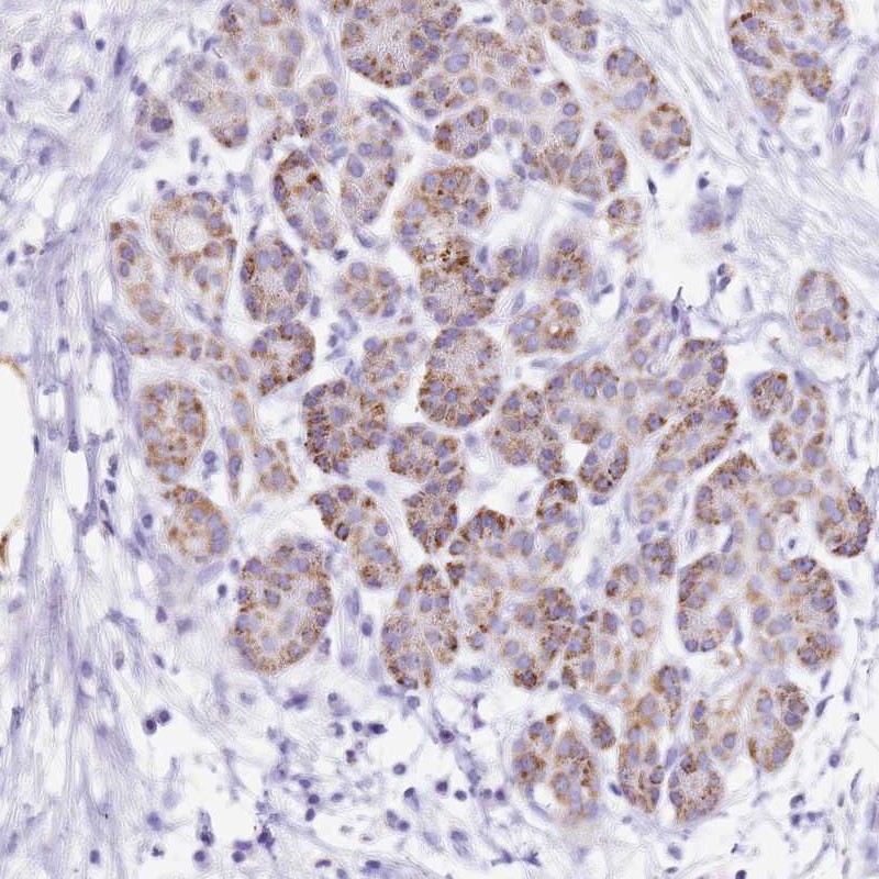
![PTBP2 antibody [C1C3] detects PTBP2 protein at nucleus on mouse middle brain by immunohistochemical analysis. Sample: Paraffin-embedded mouse middle brain. PTBP2 antibody [C1C3] (GTX116703) dilution: 1:1000.
Antigen Retrieval: Trilogy? (EDTA based, pH 8.0) buffer, 15min](https://www.genetex.com/upload/website/prouct_img/normal/GTX116703/GTX116703_40275_IHC_M_w_23060519_994.webp)
Product group Antibodies
PTBP2 antibody [C1C3]GTX116703
ApplicationsWestern Blot, ImmunoHistoChemistry, ImmunoHistoChemistry Paraffin
ReactivityHuman, Mouse, Rat
TargetPTBP2
- SizePrice
Product group Antibodies
Anti-PTBP2 Antibody144-06054
ApplicationsWestern Blot
ReactivityHuman, Mouse, Rat
TargetPTBP2
- SizePrice