

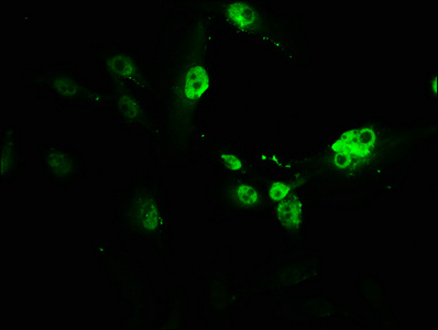
Immunofluorescence staining of U251 cells with CSB-PA019092HA01HU at 1:166, counter-stained with DAPI. The cells were fixed in 4% formaldehyde, permeabilized using 0.2% Triton X-100 and blocked in 10% normal Goat Serum. The cells were then incubated with the antibody overnight at 4°C. The secondary antibody was Alexa Fluor 488-congugated AffiniPure Goat Anti-Rabbit IgG(H+L).

")


Immunofluorescence staining of U251 cells with CSB-PA019092HA01HU at 1:166, counter-stained with DAPI. The cells were fixed in 4% formaldehyde, permeabilized using 0.2% Triton X-100 and blocked in 10% normal Goat Serum. The cells were then incubated with the antibody overnight at 4°C. The secondary antibody was Alexa Fluor 488-congugated AffiniPure Goat Anti-Rabbit IgG(H+L).
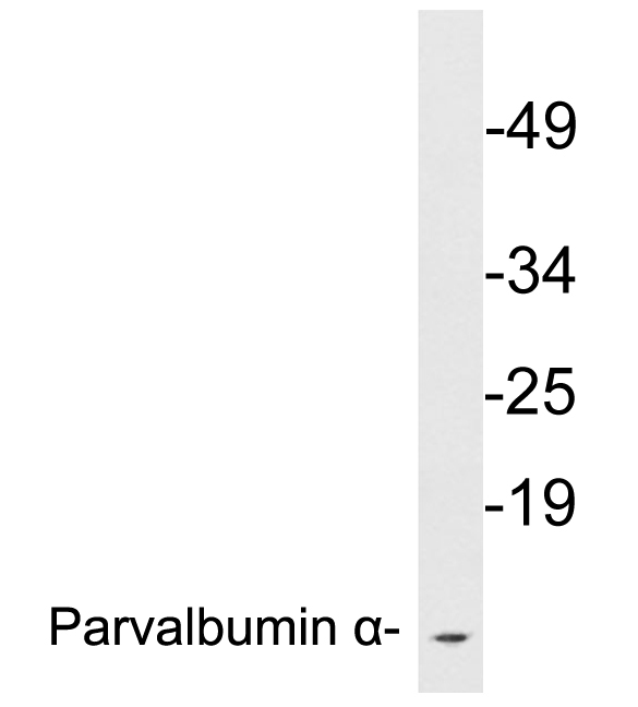
PVALB Antibody
CSB-PA019092HA01HU
ApplicationsImmunoFluorescence, Western Blot, ELISA, ImmunoHistoChemistry
Product group Antibodies
ReactivityHuman, Mouse
TargetPVALB
Overview
- SupplierCusabio
- Product NamePVALB Antibody
- Delivery Days Customer20
- ApplicationsImmunoFluorescence, Western Blot, ELISA, ImmunoHistoChemistry
- CertificationResearch Use Only
- ClonalityPolyclonal
- ConjugateUnconjugated
- Gene ID5816
- Target namePVALB
- Target descriptionparvalbumin
- Target synonymsD22S749, parvalbumin alpha, alpha-PV, alpha-parvalbumin
- HostRabbit
- IsotypeIgG
- Protein IDP20472
- Protein NameParvalbumin alpha
- Scientific DescriptionIn muscle, parvalbumin is thought to be involved in relaxation after contraction. It binds two calcium ions.
- ReactivityHuman, Mouse
- Storage Instruction-20°C or -80°C
- UNSPSC41116161
Related products
Product group Antibodies
ApplicationsWestern Blot, ELISA
ReactivityHuman, Mouse, Rat
- SizePrice
Product group Antibodies
Anti-PVALB Antibody144-60383
ApplicationsWestern Blot, ImmunoHistoChemistry
ReactivityHuman, Mouse, Rat
TargetPVALB
- SizePrice
Product group Antibodies

Parvalbumin (PVALB) AntibodyABX214662
ApplicationsWestern Blot, ELISA, ImmunoHistoChemistry
- SizePrice
Product group Antibodies
References
Parvalbumin Polyclonal AntibodyBS-1299R
ApplicationsImmunoFluorescence, Western Blot, ELISA, ImmunoCytoChemistry
ReactivityBovine, Chicken, Equine, Human, Mouse, Porcine, Rat
TargetPVALB
- SizePrice
Product group Antibodies

Goat anti-Parvalbumin, biotinylatedEB06776-B
ApplicationsWestern Blot, ELISA
ReactivityHuman, Rat
TargetPVALB
- SizePrice
Product group Antibodies
PVALB Polyclonal AntibodyCAC13889
ApplicationsImmunoFluorescence, Western Blot, ELISA, ImmunoHistoChemistry
ReactivityMouse
TargetPVALB
- SizePrice

Product group Antibodies
ApplicationsImmunoPrecipitation, Western Blot, ImmunoHistoChemistry
ReactivityHuman, Mouse, Rat
TargetPVALB
- SizePrice
![Parvalbumin antibody detects Parvalbumin protein at cell membrane and cytoplasm by immunohistochemical analysis. Sample: Paraffin-embedded mouse eye. Green: Parvalbumin stained by Parvalbumin antibody (GTX132759) diluted at 1:250. Red: beta Tubulin 3/ Tuj1, a cytoskeleton marker, stained by beta Tubulin 3/ Tuj1 antibody [GT11710] (GTX631836) diluted at 1:500. Blue: Fluoroshield with DAPI (GTX30920). Antigen Retrieval: Citrate buffer, pH 6.0, 15 min](https://www.genetex.com/upload/website/prouct_img/normal/GTX132759/GTX132759_43586_20190531_IHC-P-FL_M_w_23060523_817.webp)
Product group Antibodies
Parvalbumin antibodyGTX132759
ApplicationsWestern Blot, ImmunoHistoChemistry, ImmunoHistoChemistry Paraffin
ReactivityHuman, Mouse
TargetPVALB
- SizePrice

Product group Antibodies
Anti-PVALB AntibodyHPA048536
ApplicationsWestern Blot, ImmunoHistoChemistry
ReactivityHuman
TargetPVALB
- SizePrice