

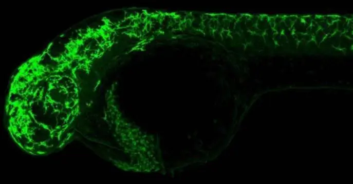
Immunohistochemical analysis (whole mount) of zebrafish embryo, using Pyruvate Dehydrogenase E1 alpha antibody (GTX104015) at 1:200 dilution.
 diluted at 1:100.")
 were separated by 10% SDS-PAGE, and the membrane was blotted with Pyruvate Dehydrogenase E1 alpha antibody (GTX104015) diluted at 1:1000. The HRP-conjugated anti-rabbit IgG antibody (GTX213110-01) was used to detect the primary antibody.")
 at 1:300 dilution.")
 A: mouse brain 10% SDS PAGE GTX104015 diluted at 1:5000 The HRP-conjugated anti-rabbit IgG antibody (GTX213110-01) was used to detect the primary antibody.")
 and Pyruvate Dehydrogenase E1 alpha knockout (KO) HeLa cell extracts (30 μg) were separated by 10% SDS-PAGE, and the membrane was blotted with Pyruvate Dehydrogenase E1 alpha antibody (GTX104015) diluted at 1:1000. The HRP-conjugated anti-rabbit IgG antibody (GTX213110-01) was used to detect the primary antibody.")
. Western blot analysis was performed using Pyruvate Dehydrogenase E1 alpha antibody (GTX104015). EasyBlot anti-Rabbit IgG (GTX221666-01) was used as a secondary reagent.")
 dilution: 1:500.
Antigen Retrieval: Trilogy? (EDTA based, pH 8.0) buffer, 15min")
 diluted at 1:2000. Blue: Fluoroshield with DAPI (GTX30920).")
 A: Rat brain 10% SDS PAGE GTX104015 diluted at 1:3000 The HRP-conjugated anti-rabbit IgG antibody (GTX213110-01) was used to detect the primary antibody.")
Immunohistochemical analysis (whole mount) of zebrafish embryo, using Pyruvate Dehydrogenase E1 alpha antibody (GTX104015) at 1:200 dilution.
Pyruvate Dehydrogenase E1 alpha antibody
GTX104015
ApplicationsImmunoFluorescence, ImmunoPrecipitation, Western Blot, ImmunoCytoChemistry, ImmunoHistoChemistry, ImmunoHistoChemistry Paraffin
Product group Antibodies
ReactivityHuman, Mouse, Rat, Zebra Fish
TargetPDHA1
Overview
- SupplierGeneTex
- Product NamePyruvate Dehydrogenase E1 alpha antibody
- Delivery Days Customer9
- Application Supplier NoteWB: 1:500-1:10000. ICC/IF: 1:100-1:1000. IHC-P: 1:100-1:1000. IP: 1:100-1:500. *Optimal dilutions/concentrations should be determined by the researcher.Not tested in other applications.
- ApplicationsImmunoFluorescence, ImmunoPrecipitation, Western Blot, ImmunoCytoChemistry, ImmunoHistoChemistry, ImmunoHistoChemistry Paraffin
- CertificationResearch Use Only
- ClonalityPolyclonal
- Concentration1.39 mg/ml
- ConjugateUnconjugated
- Gene ID5160
- Target namePDHA1
- Target descriptionpyruvate dehydrogenase E1 subunit alpha 1
- Target synonymsE1alpha, PDHA, PDHAD, PDHCE1A, PHE1A, pyruvate dehydrogenase E1 component subunit alpha, somatic form, mitochondrial, PDHE1-A type I, pyruvate dehydrogenase (lipoamide) alpha 1, pyruvate dehydrogenase E1 alpha 1 subunit, pyruvate dehydrogenase alpha 1, pyruvate dehydrogenase complex, E1-alpha polypeptide 1
- HostRabbit
- IsotypeIgG
- Protein IDP08559
- Protein NamePyruvate dehydrogenase E1 component subunit alpha, somatic form, mitochondrial
- Scientific DescriptionThe pyruvate dehydrogenase complex is a nuclear-encoded mitochondrial matrix multienzyme complex that provides the primary link between glycolysis and the tricarboxylic acid (TCA) cycle by catalyzing the irreversible conversion of pyruvate into acetyl-CoA. The PDH complex is composed of multiple copies of 3 enzymes: E1 (PDHA1); dihydrolipoyl transacetylase (DLAT; MIM 608770) (E2; EC 2.3.1.12); and dihydrolipoyl dehydrogenase (DLD; MIM 238331) (E3; EC 1.8.1.4). The E1 enzyme is a heterotetramer of 2 alpha and 2 beta subunits. The E1-alpha subunit contains the E1 active site and plays a key role in the function of the PDH complex (Brown et al., 1994 [PubMed 7853374]).[supplied by OMIM]
- ReactivityHuman, Mouse, Rat, Zebra Fish
- Storage Instruction-20°C or -80°C,2°C to 8°C
- UNSPSC41116161
Datasheet
Related products
Product group Antibodies
Anti-PDHA1 AntibodyA95816
ApplicationsWestern Blot, ELISA, ImmunoHistoChemistry
ReactivityHuman, Mouse, Rat
- SizePrice
Product group Antibodies
Anti-PDHA1 Antibody144-01895
ApplicationsImmunoFluorescence, Western Blot
ReactivityHuman, Mouse
TargetPDHA1
- SizePrice
Product group Antibodies

PDHA1 / PDH E1 Alpha AntibodyLS-C832289
ApplicationsWestern Blot, ELISA, ImmunoHistoChemistry
ReactivityHuman, Mouse, Rat
TargetPDHA1
- SizePrice
Product group Antibodies
PDHA1 Recombinant AntibodyBSM-60685R
ApplicationsImmunoFluorescence, Western Blot, ImmunoHistoChemistry, ImmunoHistoChemistry Frozen, ImmunoHistoChemistry Paraffin
ReactivityHuman, Mouse, Rat
TargetPDHA1
- SizePrice
Product group Antibodies
PDHA1 AntibodyCSB-PA003727
ApplicationsWestern Blot, ELISA, ImmunoHistoChemistry
ReactivityHuman, Mouse, Rat
TargetPDHA1
- SizePrice
Product group Antibodies
PDHA1 Polyclonal AntibodyCAC13152
ApplicationsImmunoFluorescence, ELISA, ImmunoHistoChemistry
TargetPDHA1
- SizePrice

Product group Antibodies
ApplicationsFlow Cytometry, ImmunoFluorescence, ImmunoPrecipitation, Western Blot, ImmunoCytoChemistry, ImmunoHistoChemistry
ReactivityHuman, Mouse, Rat
TargetPDHA1
- SizePrice
![Untreated (–) and treated (+) 293T whole cell extracts (30 μg) were separated by 10% SDS-PAGE, and the membrane was blotted with Pyruvate Dehydrogenase E1 alpha (phospho Ser293) antibody [GT1301] (GTX03213) diluted at 1:500. The HRP-conjugated anti-rabbit IgG antibody (GTX213110-01) was used to detect the primary antibody.](https://www.genetex.com/upload/website/prouct_img/normal/GTX03213/GTX03213_4000001567_20210716_WB_CIP_w_23053123_870.webp)
Product group Antibodies
ApplicationsWestern Blot
ReactivityHuman, Mouse, Rat
TargetPDHA1
- SizePrice

Product group Antibodies
Anti-PDHA1 AntibodyHPA063053
ApplicationsImmunoHistoChemistry
ReactivityHuman
TargetPDHA1
- SizePrice