

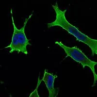
ICC/IF analysis of LOVO cells using GTX82800 Rab10 antibody [4E2]. Green : Rab10 Blue: DRAQ5 fluorescent DNA dye
![WB analysis of HeLa (1) and NIH3T3 (2) cell lysate using GTX82800 Rab10 antibody [4E2].](https://www.genetex.com/upload/website/prouct_img/normal/GTX82800/GTX82800_20170912_WB_w_23061322_713.webp "WB analysis of HeLa (1) and NIH3T3 (2) cell lysate using GTX82800 Rab10 antibody [4E2].")
ICC/IF analysis of LOVO cells using GTX82800 Rab10 antibody [4E2]. Green : Rab10 Blue: DRAQ5 fluorescent DNA dye
Rab10 antibody [4E2]
GTX82800
ApplicationsImmunoFluorescence, Western Blot, ELISA, ImmunoCytoChemistry
Product group Antibodies
ReactivityHuman, Mouse
TargetRAB10
Overview
- SupplierGeneTex
- Product NameRab10 antibody [4E2]
- Delivery Days Customer9
- Application Supplier NoteWB: 1/500 - 1/2000. ICC/IF: 1/200 - 1/1000. ELISA: 1/10000. *Optimal dilutions/concentrations should be determined by the researcher.Not tested in other applications.
- ApplicationsImmunoFluorescence, Western Blot, ELISA, ImmunoCytoChemistry
- CertificationResearch Use Only
- ClonalityMonoclonal
- Clone ID400
- ConjugateUnconjugated
- Gene ID10890
- Target nameRAB10
- Target descriptionRAB10, member RAS oncogene family
- Target synonymsras-related protein Rab-10, GTP-binding protein RAB10, ras-related GTP-binding protein
- HostMouse
- IsotypeIgG1
- Protein IDP61026
- Protein NameRas-related protein Rab-10
- Scientific DescriptionRAB10 belongs to the RAS (see HRAS; MIM 190020) superfamily of small GTPases. RAB proteins localize to exocytic and endocytic compartments and regulate intracellular vesicle trafficking (Bao et al., 1998 [PubMed 9918381]).[supplied by OMIM, Mar 2009]
- ReactivityHuman, Mouse
- Storage Instruction-20°C or -80°C,2°C to 8°C
- UNSPSC41116161
References
- Direct lysosome-based autophagy of lipid droplets in hepatocytes. Schulze RJ et al., 2020 Dec 22, Proc Natl Acad Sci U S ARead this paper
- IKKbeta activation promotes amphisome formation and extracellular vesicle secretion in tumor cells. Peng X et al., 2021 Jan, Biochim Biophys Acta Mol Cell ResRead this paper
- Maturation of Lipophagic Organelles in Hepatocytes Is Dependent Upon a Rab10/Dynamin-2 Complex. Li Z et al., 2020 Aug, HepatologyRead this paper
- A novel Rab10-EHBP1-EHD2 complex essential for the autophagic engulfment of lipid droplets. Li Z et al., 2016 Dec, Sci AdvRead this paper
Datasheet
Related products
Product group Antibodies
RAB10 AntibodyCSB-PA019152LA01HU
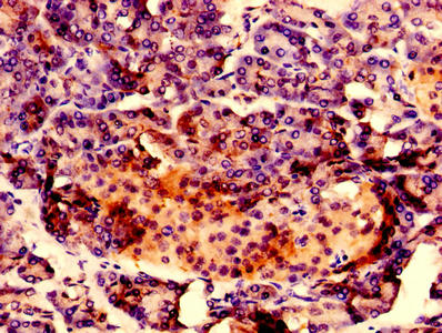
ApplicationsImmunoFluorescence, ELISA, ImmunoHistoChemistry
ReactivityHuman
TargetRAB10
- SizePrice
Product group Antibodies
Anti-RAB10 AntibodyA121629
ApplicationsWestern Blot
ReactivityCanine, Human, Monkey, Mouse, Rat
- SizePrice
Product group Antibodies

RAB10 Antibody (Biotin)LS-C316789
ApplicationsWestern Blot, ELISA
ReactivityHuman
TargetRAB10
- SizePrice

Product group Antibodies
Anti-RAB10 Antibody Picoband(r)PB9788-CARRIER-FREE
ApplicationsWestern Blot
ReactivityHamster, Human, Mouse, Rat
TargetRAB10
- SizePrice
Product group Antibodies
Anti-RAB10 Antibody144-65809
ApplicationsWestern Blot
ReactivityHuman
TargetRAB10
- SizePrice