

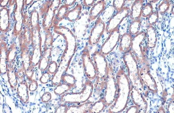
RAB35 antibody [N1C3] detects RAB35 protein at cytoplasm by immunohistochemical analysis. Sample: Paraffin-embedded rat kidney. RAB35 stained by RAB35 antibody [N1C3] (GTX120294) diluted at 1:500. Antigen Retrieval: Citrate buffer, pH 6.0, 15 min
![RAB35 antibody [N1C3] detects RAB35 protein at cytoplasm by immunohistochemical analysis. Sample: Paraffin-embedded mouse kidney. RAB35 stained by RAB35 antibody [N1C3] (GTX120294) diluted at 1:500. Antigen Retrieval: Citrate buffer, pH 6.0, 15 min](https://www.genetex.com/upload/website/prouct_img/normal/GTX120294/GTX120294_43117_20220729_IHC-P_M_22080119_192.webp "RAB35 antibody [N1C3] detects RAB35 protein at cytoplasm by immunohistochemical analysis. Sample: Paraffin-embedded mouse kidney. RAB35 stained by RAB35 antibody [N1C3] (GTX120294) diluted at 1:500. Antigen Retrieval: Citrate buffer, pH 6.0, 15 min")
 antibody at 1:250 dilution.
Antigen Retrieval: Citrate buffer, pH 6.0, 15 min")
![RAB35 antibody [N1C3] detects RAB35 protein at Golgi apparatus by immunofluorescent analysis. Sample: U87-MG cells were fixed in 4% paraformaldehyde at RT for 15 min. Green: RAB35 stained by RAB35 antibody [N1C3] (GTX120294) diluted at 1:500. Blue: Hoechst 33342 staining.](https://www.genetex.com/upload/website/prouct_img/normal/GTX120294/GTX120294_43117_20190424_ICC_IF_w_23060519_220.webp "RAB35 antibody [N1C3] detects RAB35 protein at Golgi apparatus by immunofluorescent analysis. Sample: U87-MG cells were fixed in 4% paraformaldehyde at RT for 15 min. Green: RAB35 stained by RAB35 antibody [N1C3] (GTX120294) diluted at 1:500. Blue: Hoechst 33342 staining.")
![Various whole cell extracts (30 μg) were separated by 12% SDS-PAGE, and the membrane was blotted with RAB35 antibody [N1C3] (GTX120294) diluted at 1:1000. The HRP-conjugated anti-rabbit IgG antibody (GTX213110-01) was used to detect the primary antibody.](https://www.genetex.com/upload/website/prouct_img/normal/GTX120294/GTX120294_43040_20190823_WB_w_23060519_361.webp "Various whole cell extracts (30 μg) were separated by 12% SDS-PAGE, and the membrane was blotted with RAB35 antibody [N1C3] (GTX120294) diluted at 1:1000. The HRP-conjugated anti-rabbit IgG antibody (GTX213110-01) was used to detect the primary antibody.")
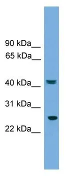
![Mouse tissue extract (50 μg) was separated by 12% SDS-PAGE, and the membrane was blotted with RAB35 antibody [N1C3] (GTX120294) diluted at 1:1000. The HRP-conjugated anti-rabbit IgG antibody (GTX213110-01) was used to detect the primary antibody.](https://www.genetex.com/upload/website/prouct_img/normal/GTX120294/GTX120294_43117_20180216_WB_M_brain_w_23060519_977.webp "Mouse tissue extract (50 μg) was separated by 12% SDS-PAGE, and the membrane was blotted with RAB35 antibody [N1C3] (GTX120294) diluted at 1:1000. The HRP-conjugated anti-rabbit IgG antibody (GTX213110-01) was used to detect the primary antibody.")
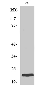
![Non-transfected (–) and transfected (+) 293T whole cell extracts (30 μg) were separated by 12% SDS-PAGE, and the membrane was blotted with RAB35 antibody [N1C3] (GTX120294) diluted at 1:2000. The HRP-conjugated anti-rabbit IgG antibody (GTX213110-01) was used to detect the primary antibody.](https://www.genetex.com/upload/website/prouct_img/normal/GTX120294/GTX120294_43040_20190823_WB_B_w_23060519_524.webp "Non-transfected (–) and transfected (+) 293T whole cell extracts (30 μg) were separated by 12% SDS-PAGE, and the membrane was blotted with RAB35 antibody [N1C3] (GTX120294) diluted at 1:2000. The HRP-conjugated anti-rabbit IgG antibody (GTX213110-01) was used to detect the primary antibody.")
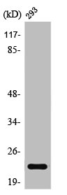
![Whole cell extract (30 μg) was separated by 12% SDS-PAGE, and the membrane was blotted with RAB35 antibody [N1C3] (GTX120294) diluted at 1:1000. The HRP-conjugated anti-rabbit IgG antibody (GTX213110-01) was used to detect the primary antibody.](https://www.genetex.com/upload/website/prouct_img/normal/GTX120294/GTX120294_43117_20231222_WB_23122619_120.webp "Whole cell extract (30 μg) was separated by 12% SDS-PAGE, and the membrane was blotted with RAB35 antibody [N1C3] (GTX120294) diluted at 1:1000. The HRP-conjugated anti-rabbit IgG antibody (GTX213110-01) was used to detect the primary antibody.")
RAB35 antibody [N1C3] detects RAB35 protein at cytoplasm by immunohistochemical analysis. Sample: Paraffin-embedded rat kidney. RAB35 stained by RAB35 antibody [N1C3] (GTX120294) diluted at 1:500. Antigen Retrieval: Citrate buffer, pH 6.0, 15 min
RAB35 antibody [N1C3]
GTX120294
ApplicationsImmunoFluorescence, Western Blot, ImmunoCytoChemistry, ImmunoHistoChemistry, ImmunoHistoChemistry Paraffin
Product group Antibodies
ReactivityHamster, Human, Mouse, Rat
TargetRAB35
Overview
- SupplierGeneTex
- Product NameRAB35 antibody [N1C3]
- Delivery Days Customer9
- Application Supplier NoteWB: 1:500-1:3000. ICC/IF: 1:100-1:1000. IHC-P: 1:100-1:1000. *Optimal dilutions/concentrations should be determined by the researcher.Not tested in other applications.
- ApplicationsImmunoFluorescence, Western Blot, ImmunoCytoChemistry, ImmunoHistoChemistry, ImmunoHistoChemistry Paraffin
- CertificationResearch Use Only
- ClonalityPolyclonal
- Concentration0.24 mg/ml
- ConjugateUnconjugated
- Gene ID11021
- Target nameRAB35
- Target descriptionRAB35, member RAS oncogene family
- Target synonymsH-ray, RAB1C, RAY, ras-related protein Rab-35, GTP-binding protein RAY, ras-related protein rab-1c (GTP-binding protein ray)
- HostRabbit
- IsotypeIgG
- Protein IDQ15286
- Protein NameRas-related protein Rab-35
- ReactivityHamster, Human, Mouse, Rat
- Storage Instruction-20°C or -80°C,2°C to 8°C
- UNSPSC41116161
Datasheet
Related products
Product group Antibodies
Anti-RAB35 AntibodyA97312
ApplicationsWestern Blot, ELISA
ReactivityHuman, Mouse, Rat
- SizePrice
Product group Antibodies
Anti-RAB35 (C-term) Antibody102-23869
ApplicationsWestern Blot, ImmunoHistoChemistry, ImmunoHistoChemistry Paraffin
TargetRAB35
- SizePrice

Product group Antibodies
Anti-RAB35 Antibody (C-term)A03845-1
ApplicationsWestern Blot, ImmunoHistoChemistry, ImmunoHistoChemistry Paraffin
ReactivityHuman, Mouse, Rat
TargetRAB35
- SizePrice
Product group Antibodies
RAB35 Polyclonal AntibodyBS-7765R
ApplicationsFlow Cytometry, ImmunoFluorescence, ELISA, ImmunoCytoChemistry, ImmunoHistoChemistry, ImmunoHistoChemistry Frozen, ImmunoHistoChemistry Paraffin
ReactivityBovine, Canine, Chicken, Equine, Human, Rabbit
TargetRAB35
- SizePrice
Product group Antibodies
RAB35 AntibodyCSB-PA003896
ApplicationsWestern Blot, ELISA
ReactivityHuman, Mouse, Rat
TargetRAB35
- SizePrice
Product group Antibodies
Rab35 Polyclonal AntibodyCAC09264
ApplicationsImmunoFluorescence, ELISA, ImmunoHistoChemistry
TargetRAB35
- SizePrice
Product group Antibodies

RAB35 AntibodyLS-C402107
ApplicationsELISA, ImmunoHistoChemistry
ReactivityHuman, Mouse, Rat
TargetRAB35
- SizePrice

Product group Antibodies
Anti-RAB35 AntibodyHPA054146
ApplicationsImmunoHistoChemistry
ReactivityHuman
TargetRAB35
- SizePrice
Product group Antibodies
References
RAB35 antibodyGTX35180
ApplicationsWestern Blot
ReactivityHuman
TargetRAB35
- SizePrice