

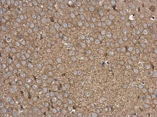
Rabphilin 3A antibody detects Rabphilin 3A protein at cell membrane and cytoplasm in rat brain by immunohistochemical analysis. Sample: Paraffin-embedded rat brain. Rabphilin 3A antibody (GTX115646) diluted at 1:500.
Antigen Retrieval: Citrate buffer, pH 6.0, 15 min
![Rabphilin 3A antibody detects Rabphilin 3A protein by immunofluorescent analysis. Sample: DIV9 rat E18 primary cortical neuron cells were fixed in 4% paraformaldehyde at RT for 15 min. Green: Rabphilin 3A stained by Rabphilin 3A antibody (GTX115646) diluted at 1:500. Red: beta Tubulin 3/ Tuj1, stained by beta Tubulin 3/ Tuj1 antibody [GT1338] (GTX631831) diluted at 1:500. Blue: Fluoroshield with DAPI (GTX30920).](https://www.genetex.com/upload/website/prouct_img/normal/GTX115646/GTX115646_41269_20171115_ICC_IF_R_w_23060519_213.webp "Rabphilin 3A antibody detects Rabphilin 3A protein by immunofluorescent analysis. Sample: DIV9 rat E18 primary cortical neuron cells were fixed in 4% paraformaldehyde at RT for 15 min. Green: Rabphilin 3A stained by Rabphilin 3A antibody (GTX115646) diluted at 1:500. Red: beta Tubulin 3/ Tuj1, stained by beta Tubulin 3/ Tuj1 antibody [GT1338] (GTX631831) diluted at 1:500. Blue: Fluoroshield with DAPI (GTX30920).")
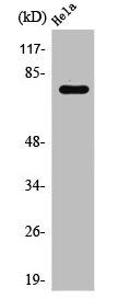
 were separated by 7.5% SDS-PAGE, and the membrane was blotted with Rabphilin 3A antibody (GTX115646) diluted at 1:3000. The HRP-conjugated anti-rabbit IgG antibody (GTX213110-01) was used to detect the primary antibody, and the signal was developed with Trident ECL plus-Enhanced.")
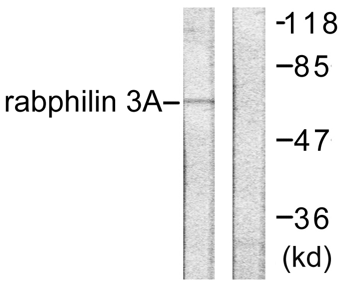
 was separated by 7.5% SDS-PAGE, and the membrane was blotted with Rabphilin 3A antibody (GTX115646) diluted at 1:1000. The HRP-conjugated anti-rabbit IgG antibody (GTX213110-01) was used to detect the primary antibody.")
 dilution: 1:500.
Antigen Retrieval: Trilogy? (EDTA based, pH 8.0) buffer, 15min")
Rabphilin 3A antibody detects Rabphilin 3A protein at cell membrane and cytoplasm in rat brain by immunohistochemical analysis. Sample: Paraffin-embedded rat brain. Rabphilin 3A antibody (GTX115646) diluted at 1:500.
Antigen Retrieval: Citrate buffer, pH 6.0, 15 min
Rabphilin 3A antibody
GTX115646
ApplicationsImmunoFluorescence, Western Blot, ImmunoCytoChemistry, ImmunoHistoChemistry, ImmunoHistoChemistry Paraffin
Product group Antibodies
ReactivityHuman, Mouse, Rat
TargetRPH3A
Overview
- SupplierGeneTex
- Product NameRabphilin 3A antibody
- Delivery Days Customer9
- Application Supplier NoteWB: 1:500-1:3000. ICC/IF: 1:100-1:1000. IHC-P: 1:100-1:1000. *Optimal dilutions/concentrations should be determined by the researcher.Not tested in other applications.
- ApplicationsImmunoFluorescence, Western Blot, ImmunoCytoChemistry, ImmunoHistoChemistry, ImmunoHistoChemistry Paraffin
- CertificationResearch Use Only
- ClonalityPolyclonal
- Concentration1 mg/ml
- ConjugateUnconjugated
- Gene ID22895
- Target nameRPH3A
- Target descriptionrabphilin 3A
- Target synonymsrabphilin-3A, exophilin-1, rabphilin 3A homolog
- HostRabbit
- IsotypeIgG
- Protein IDQ9Y2J0
- Protein NameRabphilin-3A
- Scientific DescriptionExocytosis of neurotransmitters and hormones is fundamental to synaptic neurotransmission and cell-cell communication. RAB3A (MIM 179390) is a small G protein that is thought to act at late stages of exocytosis, and RPH3A is a RAB3A effector (Lin et al., 2007 [PubMed 17149709]).[supplied by OMIM]
- ReactivityHuman, Mouse, Rat
- Storage Instruction-20°C or -80°C,2°C to 8°C
- UNSPSC41116161
Datasheet
Related products
Product group Antibodies
RPH3A AntibodyCSB-PA003905
ApplicationsImmunoFluorescence, Western Blot, ELISA, ImmunoHistoChemistry
ReactivityHuman, Mouse, Rat
TargetRPH3A
- SizePrice

Product group Antibodies
Anti-RPH3A Antibody Picoband(r)A10247-2-CARRIER-FREE
ApplicationsWestern Blot, ELISA, ImmunoHistoChemistry
ReactivityHuman, Mouse, Rat
TargetRPH3A
- SizePrice
Product group Antibodies
ApplicationsImmunoFluorescence, Western Blot, ELISA, ImmunoHistoChemistry
ReactivityHuman, Mouse, Rat
- SizePrice

Product group Antibodies
Anti-RPH3A AntibodyHPA002475
ApplicationsImmunoHistoChemistry
ReactivityHuman
TargetRPH3A
- SizePrice
Product group Antibodies

RPH3A / Rabphilin 3A Antibody (Internal)LS-C368643
ApplicationsImmunoFluorescence, Western Blot, ImmunoCytoChemistry, ImmunoHistoChemistry, ImmunoHistoChemistry Paraffin
ReactivityHuman, Mouse, Rat
TargetRPH3A
- SizePrice
Product group Antibodies
Rabphilin 3A antibodyGTX33452
ApplicationsWestern Blot
ReactivityHuman, Mouse, Rat
TargetRPH3A
- SizePrice
Product group Antibodies
Anti-RPH3A Antibody144-06722
ApplicationsWestern Blot
ReactivityHuman, Mouse, Rat
TargetRPH3A
- SizePrice