

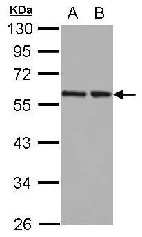
![Western blot using GeneTex's affinity purified anti-HR23B antibody shows detection of a band at ~58 kDa (arrowhead) corresponding to HR23B present in a HeLa whole cell lysate. Pre-incubation of antibody with immunizing peptide completely blocks reactivity (data not shown). Approximately 33 μg of lysate was separated by 4-20% Tris Glycine SDS-PAGE. After blocking the membrane was probed overnight at 4oC with the primary antibody diluted to 1:500 in 5% BLOTTO in PBS. The membrane was washed and reacted with a 1:20,000 dilution of IRDye800 conjugated rabbit anti-Goat IgG [H&L] for 45 min at room temperature (800 nm channel, green). Molecular weight estimation was made by comparison to prestained MW markers indicated at left (700 nm channel, red). IRDye800 fluorescence image was captured using the OdysseyR Infrared Imaging System developed by LI-COR. IRDye is a trademark of LI-COR, Inc. Other detection systems will yield similar results.](https://www.genetex.com/upload/website/prouct_img/normal/GTX48751/GTX48751_20160330_WB_w_23060823_782.webp "Western blot using GeneTex's affinity purified anti-HR23B antibody shows detection of a band at ~58 kDa (arrowhead) corresponding to HR23B present in a HeLa whole cell lysate. Pre-incubation of antibody with immunizing peptide completely blocks reactivity (data not shown). Approximately 33 μg of lysate was separated by 4-20% Tris Glycine SDS-PAGE. After blocking the membrane was probed overnight at 4oC with the primary antibody diluted to 1:500 in 5% BLOTTO in PBS. The membrane was washed and reacted with a 1:20,000 dilution of IRDye800 conjugated rabbit anti-Goat IgG [H&L] for 45 min at room temperature (800 nm channel, green). Molecular weight estimation was made by comparison to prestained MW markers indicated at left (700 nm channel, red). IRDye800 fluorescence image was captured using the OdysseyR Infrared Imaging System developed by LI-COR. IRDye is a trademark of LI-COR, Inc. Other detection systems will yield similar results.")

RAD23B antibody
GTX48751
ApplicationsWestern Blot, ELISA
Product group Antibodies
ReactivityHuman
TargetRAD23B
Overview
- SupplierGeneTex
- Product NameRAD23B antibody
- Delivery Days Customer9
- Application Supplier NoteWB: 1:500-1:2000. ELISA: 1:2000-1:10000. *Optimal dilutions/concentrations should be determined by the researcher.Not tested in other applications.
- ApplicationsWestern Blot, ELISA
- CertificationResearch Use Only
- ClonalityPolyclonal
- Concentration1.1 mg/ml
- ConjugateUnconjugated
- Gene ID5887
- Target nameRAD23B
- Target descriptionRAD23 nucleotide excision repair protein B
- Target synonymsHHR23B, HR23B, P58, UV excision repair protein RAD23 homolog B, RAD23 homolog B, nucleotide excision repair protein, RAD23, yeast homolog of, B, XP-C repair complementing complex 58 kDa, XP-C repair complementing protein, XP-C repair-complementing complex 58 kDa protein
- HostGoat
- IsotypeIgG
- Protein IDP54727
- Protein NameUV excision repair protein RAD23 homolog B
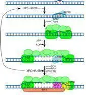
- Scientific DescriptionHR23B (also known as UV excision repair protein RAD23 homolog B, XP-C repair complementing complex 58 kDa protein and p58) is one of two human homologs of Saccharomyces cerevisiae Rad23 (hHR23A and hHR23B), a protein involved in nucleotide excision repair (NER). This protein was shown to interact with, and elevate the nucleotide excision activity of 3-methyladenine-DNA glycosylase (MPG), which suggested a role in DNA damage recognition in base excision repair. This protein contains an N-terminal ubiquitin-like domain, which was reported to interact with 26S proteasome, as well as with ubiquitin protein ligase E6AP, and thus suggests that this protein may be involved in the ubiquitin mediated proteolytic pathway in cells.
- ReactivityHuman
- Storage Instruction-20°C or -80°C,2°C to 8°C
- UNSPSC41116161
Datasheet
Related products
Product group Antibodies
RAD23B AntibodyCSB-PA003914
ApplicationsImmunoFluorescence, Western Blot, ELISA, ImmunoHistoChemistry
ReactivityHuman, Mouse, Rat
TargetRAD23B
- SizePrice

Product group Antibodies
Anti-RAD23B AntibodyA96758
ApplicationsWestern Blot, ELISA
ReactivityHuman, Mouse, Rat
- SizePrice
Product group Antibodies

Anti-RAD23B [SAIC-28A-29]Ab00320-1.1
ApplicationsMass Spectrometry, Western Blot
ReactivityHuman
TargetRAD23B
- SizePrice
Product group Antibodies

RAD23B / HR23B AntibodyLS-C832013
ApplicationsWestern Blot, ELISA, ImmunoHistoChemistry
ReactivityHuman, Rat
TargetRAD23B
- SizePrice

Product group Antibodies
Anti-RAD23B AntibodyHPA029718
ApplicationsWestern Blot, ImmunoCytoChemistry, ImmunoHistoChemistry
ReactivityHuman
TargetRAD23B
- SizePrice
Product group Antibodies
RAD23B Polyclonal AntibodyCAC13894
ApplicationsWestern Blot, ELISA, ImmunoHistoChemistry
TargetRAD23B
- SizePrice
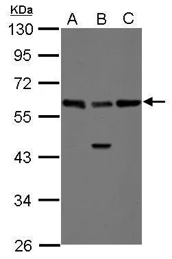
![WB analysis of K562, SW480, CHO-K1, 3T3, and COS7 cell lysates using GTX66845 RAD23B antibody [5H1]. Dilution : 1:1000](https://www.genetex.com/upload/website/prouct_img/normal/GTX66845/GTX66845_20190305_WB_w_23061221_284.webp)
Product group Antibodies
RAD23B antibody [5H1]GTX66845
ApplicationsImmunoFluorescence, Western Blot, ImmunoCytoChemistry, ImmunoHistoChemistry, ImmunoHistoChemistry Paraffin
ReactivityHamster, Human, Monkey, Mouse
TargetRAD23B
- SizePrice
Product group Antibodies
RAD23B antibodyGTX103424
ApplicationsImmunoFluorescence, Western Blot, ImmunoCytoChemistry, ImmunoHistoChemistry, ImmunoHistoChemistry Paraffin
ReactivityHuman
TargetRAD23B
- SizePrice
Product group Antibodies
RAD23B antibodyGTX113820
ApplicationsImmunoFluorescence, Western Blot, ImmunoCytoChemistry, ImmunoHistoChemistry, ImmunoHistoChemistry Paraffin
ReactivityHuman
TargetRAD23B
- SizePrice
Product group Antibodies
RAD23B antibodyGTX85831
ApplicationsWestern Blot, ELISA, ImmunoHistoChemistry, ImmunoHistoChemistry Paraffin
ReactivityHuman
TargetRAD23B
- SizePrice