


C57BL/6 mouse bone marrow cells were stained with Rat Anti-Mouse CD106-FITC (SB Cat. 1510-02) and Rat Anti-Mouse Ly-6G/Ly-6C-PE (SB Cat. No. 1900-09).
C57BL/6 mouse bone marrow cells were stained with Rat Anti-Mouse CD106-FITC (SB Cat. 1510-02) and Rat Anti-Mouse Ly-6G/Ly-6C-PE (SB Cat. No. 1900-09).
Rat Anti-Mouse CD106-FITC
1510-02
ApplicationsFlow Cytometry, Western Blot, ImmunoHistoChemistry, ImmunoHistoChemistry Frozen
Product group Antibodies
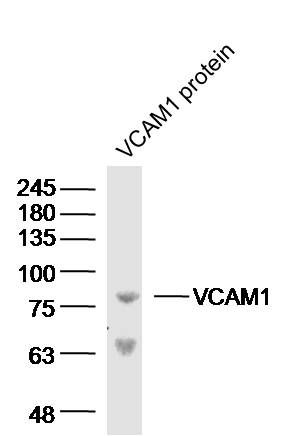
TargetVcam1
Overview
- SupplierSouthernBiotech
- Product NameRat Anti-Mouse CD106-FITC
- Delivery Days Customer7
- ApplicationsFlow Cytometry, Western Blot, ImmunoHistoChemistry, ImmunoHistoChemistry Frozen
- CertificationResearch Use Only
- ClonalityMonoclonal
- Clone IDM/K-2
- ConjugateFITC
- Gene ID22329
- Target nameVcam1
- Target descriptionvascular cell adhesion molecule 1
- Target synonymsCD106; vascular cell adhesion protein 1; Vcam; V-CAM 1; Vcam-1
- HostRat
- IsotypeIgG1
- Protein IDP29533
- Protein NameVascular cell adhesion protein 1
- Storage Instruction2°C to 8°C
- UNSPSC12352203
Related products
Product group Antibodies
References
VCAM1 Polyclonal AntibodyBS-0396R
ApplicationsFlow Cytometry, ImmunoFluorescence, Western Blot, ELISA, ImmunoCytoChemistry, ImmunoHistoChemistry, ImmunoHistoChemistry Frozen, ImmunoHistoChemistry Paraffin
TargetVcam1
- SizePrice
Product group Antibodies
References
VCAM1 / CD106 antibody [M/K-2.7]GTX14360
ApplicationsImmunoFluorescence, ImmunoCytoChemistry, Neutralisation/Blocking
TargetVcam1
- SizePrice

Product group Antibodies
Anti-VCAM1 Antibody Picoband(r)A01199-CARRIER-FREE
ApplicationsWestern Blot, ELISA, ImmunoHistoChemistry
TargetVcam1
- SizePrice