

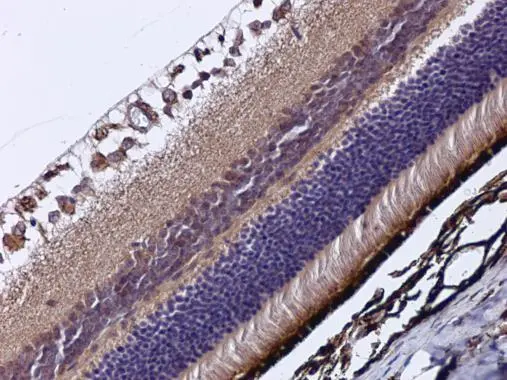
RBP3 antibody [C2C3], C-term detects RBP3 protein at cytoplasm in mouse eye by immunohistochemical analysis. Sample: Paraffin-embedded mouse eye. RBP3 antibody [C2C3], C-term (GTX114714) diluted at 1:500.
Antigen Retrieval: Citrate buffer, pH 6.0, 15 min
![RBP3 antibody [C2C3], C-term detects RBP3 protein at cytoplasm by immunofluorescent analysis. Sample: HeLa cells were fixed in ice-cold MeOH for 5 min. Green: RBP3 protein stained by RBP3 antibody [C2C3], C-term (GTX114714) diluted at 1:400. Blue: Hoechst 33342 staining.](https://www.genetex.com/upload/website/prouct_img/normal/GTX114714/GTX114714_40219_20151030_IFA_w_23060518_249.webp "RBP3 antibody [C2C3], C-term detects RBP3 protein at cytoplasm by immunofluorescent analysis. Sample: HeLa cells were fixed in ice-cold MeOH for 5 min. Green: RBP3 protein stained by RBP3 antibody [C2C3], C-term (GTX114714) diluted at 1:400. Blue: Hoechst 33342 staining.")
RBP3 antibody [C2C3], C-term detects RBP3 protein at cytoplasm in mouse eye by immunohistochemical analysis. Sample: Paraffin-embedded mouse eye. RBP3 antibody [C2C3], C-term (GTX114714) diluted at 1:500.
Antigen Retrieval: Citrate buffer, pH 6.0, 15 min
RBP3 antibody [C2C3], C-term
GTX114714
ApplicationsImmunoFluorescence, ImmunoCytoChemistry, ImmunoHistoChemistry, ImmunoHistoChemistry Paraffin
Product group Antibodies
ReactivityHuman, Mouse
TargetRBP3
Overview
- SupplierGeneTex
- Product NameRBP3 antibody [C2C3], C-term
- Delivery Days Customer9
- Application Supplier NoteICC/IF: 1:100-1:1000. IHC-P: 1:100-1:1000. *Optimal dilutions/concentrations should be determined by the researcher.Not tested in other applications.
- ApplicationsImmunoFluorescence, ImmunoCytoChemistry, ImmunoHistoChemistry, ImmunoHistoChemistry Paraffin
- CertificationResearch Use Only
- ClonalityPolyclonal
- Concentration0.56 mg/ml
- ConjugateUnconjugated
- Gene ID5949
- Target nameRBP3
- Target descriptionretinol binding protein 3
- Target synonymsD10S64, D10S65, D10S66, IRBP, RBPI, RP66, retinol-binding protein 3, interphotoreceptor retinoid-binding protein, interstitial retinol binding protein 3, retinol binding protein 3, interstitial
- HostRabbit
- IsotypeIgG
- Protein IDP10745
- Protein NameRetinol-binding protein 3
- Scientific DescriptionInterphotoreceptor retinol-binding protein is a large glycoprotein known to bind retinoids and found primarily in the interphotoreceptor matrix of the retina between the retinal pigment epithelium and the photoreceptor cells. It is thought to transport retinoids between the retinal pigment epithelium and the photoreceptors, a critical role in the visual process.The human IRBP gene is approximately 9.5 kbp in length and consists of four exons separated by three introns. The introns are 1.6-1.9 kbp long. The gene is transcribed by photoreceptor and retinoblastoma cells into an approximately 4.3-kilobase mRNA that is translated and processed into a glycosylated protein of 135,000 Da. The amino acid sequence of human IRBP can be divided into four contiguous homology domains with 33-38% identity, suggesting a series of gene duplication events. In the gene, the boundaries of these domains are not defined by exon-intron junctions, as might have been expected. The first three homology domains and part of the fourth are all encoded by the first large exon, which is 3,180 base pairs long. The remainder of the fourth domain is encoded in the last three exons, which are 191, 143, and approximately 740 base pairs long, respectively. [provided by RefSeq]
- ReactivityHuman, Mouse
- Storage Instruction-20°C or -80°C,2°C to 8°C
- UNSPSC41116161
Datasheet
Related products
Product group Antibodies
Anti-RBP3 AntibodyA31357
ApplicationsImmunoFluorescence, Western Blot, ImmunoHistoChemistry
ReactivityHuman
- SizePrice

Product group Antibodies
Anti-IRBP/RBP3 Antibody Picoband(r)A02913-1-CARRIER-FREE
ApplicationsFlow Cytometry, Western Blot, ELISA
ReactivityHuman, Mouse, Rat
TargetRBP3
- SizePrice
Product group Antibodies
Anti-RBP3 Antibody144-06403
ApplicationsImmunoFluorescence, Western Blot
ReactivityHuman, Mouse, Rat
TargetRBP3
- SizePrice
Product group Antibodies

RBP3 / IRBP AntibodyLS-C831436
ApplicationsImmunoHistoChemistry
ReactivityHuman, Mouse, Rat
TargetRBP3
- SizePrice
Product group Antibodies
ApplicationsImmunoPrecipitation, Western Blot, ImmunoCytoChemistry, ImmunoHistoChemistry
ReactivityMouse, Rat
TargetRBP3
- SizePrice
Product group Antibodies
RBP3 AntibodyCSB-PA019482LA01HU
ApplicationsELISA
ReactivityHuman
TargetRBP3
- SizePrice

Product group Antibodies
Anti-RBP3 AntibodyHPA041301
ApplicationsImmunoHistoChemistry
ReactivityHuman
TargetRBP3
- SizePrice
Product group Antibodies
Anti-RBP3 AntibodyCAB6403
ApplicationsWestern Blot, ELISA
ReactivityHuman
TargetRBP3
- SizePrice