

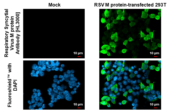
Respiratory Syncytial Virus M protein Antibody [HL3000] detects Respiratory Syncytial Virus M protein Antibody protein by immunofluorescent analysis. Sample: Mock and transfected 293T cells were fixed in ice-cold MeOH for 5 min. Green: Respiratory Syncytial Virus M Antibody stained by Respiratory Syncytial Virus M protein Antibody [HL3000] (GTX640410) diluted at 1:500. Blue: Fluoroshield with DAPI (GTX30920).
![Non-infected (–) and infected (+) HEp2 whole cell extracts were separated by 12% SDS-PAGE, and the membrane was blotted with Respiratory Syncytial Virus M protein Antibody [HL3000] (GTX640410) diluted at 1:1000. The HRP-conjugated anti-rabbit IgG antibody (GTX213110-01) was used to detect the primary antibody.](https://www.genetex.com/upload/website/prouct_img/normal/GTX640410/GTX640410_T-45418_20240628_WB_RSV_24070822_623.webp "Non-infected (–) and infected (+) HEp2 whole cell extracts were separated by 12% SDS-PAGE, and the membrane was blotted with Respiratory Syncytial Virus M protein Antibody [HL3000] (GTX640410) diluted at 1:1000. The HRP-conjugated anti-rabbit IgG antibody (GTX213110-01) was used to detect the primary antibody.")
![RSV type A and type B viral lysate were separated by 12% SDS-PAGE, and the membrane was blotted with Respiratory Syncytial Virus M protein Antibody [HL3000] (GTX640410) diluted at 1:1000. The HRP-conjugated anti-rabbit IgG antibody (GTX213110-01) was used to detect the primary antibody.](https://www.genetex.com/upload/website/prouct_img/normal/GTX640410/GTX640410_45502_20240816_WB_RSV_24082301_218.webp "RSV type A and type B viral lysate were separated by 12% SDS-PAGE, and the membrane was blotted with Respiratory Syncytial Virus M protein Antibody [HL3000] (GTX640410) diluted at 1:1000. The HRP-conjugated anti-rabbit IgG antibody (GTX213110-01) was used to detect the primary antibody.")
Respiratory Syncytial Virus M protein Antibody [HL3000] detects Respiratory Syncytial Virus M protein Antibody protein by immunofluorescent analysis. Sample: Mock and transfected 293T cells were fixed in ice-cold MeOH for 5 min. Green: Respiratory Syncytial Virus M Antibody stained by Respiratory Syncytial Virus M protein Antibody [HL3000] (GTX640410) diluted at 1:500. Blue: Fluoroshield with DAPI (GTX30920).
Respiratory Syncytial Virus M protein Antibody [HL3000]
GTX640410
ApplicationsImmunoFluorescence, Western Blot, ImmunoCytoChemistry
Product group Antibodies
ReactivityVirus
Overview
- SupplierGeneTex
- Product NameRespiratory Syncytial Virus M protein Antibody [HL3000]
- Delivery Days Customer7
- Application Supplier NoteWB: 1:500-1:3000. ICC/IF: 1:100-1:1000. *Optimal dilutions/concentrations should be determined by the researcher.Not tested in other applications.
- ApplicationsImmunoFluorescence, Western Blot, ImmunoCytoChemistry
- CertificationResearch Use Only
- ClonalityMonoclonal
- Clone IDHL3000
- Concentration1 mg/ml
- ConjugateUnconjugated
- HostRabbit
- IsotypeIgG
- ReactivityVirus
- Storage Instruction-20°C or -80°C,2°C to 8°C
- UNSPSC12352203