

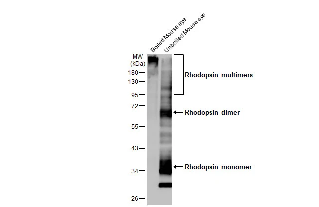
Boiled and unboiled various tissue extracts (50 μg) were separated by 10% SDS-PAGE, and the membrane was blotted with Rhodopsin antibody [HL2668] (GTX639332) diluted at 1:1000. The HRP-conjugated anti-rabbit IgG antibody (GTX213110-01) was used to detect the primary antibody.
![Rhodopsin antibody [HL2668] detects Rhodopsin protein by immunohistochemical analysis. Sample: Paraffin-embedded mouse eye. Rhodopsin stained by Rhodopsin antibody [HL2668] (GTX639332) diluted at 1:100. Red: beta Tubulin 3/ Tuj1 stained by beta Tubulin 3/ Tuj1 antibody [GT11710] (GTX631836) diluted at 1:500. Blue: Fluoroshield with DAPI (GTX30920). Antigen Retrieval: Citrate buffer, pH 6.0, 15 min](https://www.genetex.com/upload/website/prouct_img/normal/GTX639332/GTX639332_T-45215_20231201_IHC-P_M_23120519_665.webp "Rhodopsin antibody [HL2668] detects Rhodopsin protein by immunohistochemical analysis. Sample: Paraffin-embedded mouse eye. Rhodopsin stained by Rhodopsin antibody [HL2668] (GTX639332) diluted at 1:100. Red: beta Tubulin 3/ Tuj1 stained by beta Tubulin 3/ Tuj1 antibody [GT11710] (GTX631836) diluted at 1:500. Blue: Fluoroshield with DAPI (GTX30920). Antigen Retrieval: Citrate buffer, pH 6.0, 15 min")
![Rat tissue extract (30 μg) was separated by 10% SDS-PAGE, and the membrane was blotted with Rhodopsin antibody [HL2668] (GTX639332) diluted at 1:500. The HRP-conjugated anti-rabbit IgG antibody (GTX213110-01) was used to detect the primary antibody.](https://www.genetex.com/upload/website/prouct_img/normal/GTX639332/GTX639332_T-45215_20231201_WB_R_eye_23120519_478.webp "Rat tissue extract (30 μg) was separated by 10% SDS-PAGE, and the membrane was blotted with Rhodopsin antibody [HL2668] (GTX639332) diluted at 1:500. The HRP-conjugated anti-rabbit IgG antibody (GTX213110-01) was used to detect the primary antibody.")
![Rhodopsin antibody [HL2668] detects Rhodopsin protein by immunohistochemical analysis. Sample: Paraffin-embedded rat eye. Rhodopsin stained by Rhodopsin antibody [HL2668] (GTX639332) diluted at 1:100. Red: beta Tubulin 3/ Tuj1 stained by beta Tubulin 3/ Tuj1 antibody [GT11710] (GTX631836) diluted at 1:500. Blue: Fluoroshield with DAPI (GTX30920). Antigen Retrieval: Citrate buffer, pH 6.0, 15 min](https://www.genetex.com/upload/website/prouct_img/normal/GTX639332/GTX639332_T-45215_20231201_IHC-P_R_23120519_459.webp "Rhodopsin antibody [HL2668] detects Rhodopsin protein by immunohistochemical analysis. Sample: Paraffin-embedded rat eye. Rhodopsin stained by Rhodopsin antibody [HL2668] (GTX639332) diluted at 1:100. Red: beta Tubulin 3/ Tuj1 stained by beta Tubulin 3/ Tuj1 antibody [GT11710] (GTX631836) diluted at 1:500. Blue: Fluoroshield with DAPI (GTX30920). Antigen Retrieval: Citrate buffer, pH 6.0, 15 min")
![Non-transfected (–) and transfected (+) boiled and unboiled 293T whole cell extracts (30 μg) were separated by 10% SDS-PAGE, and the membrane was blotted with Rhodopsin antibody [HL2668] (GTX639332) diluted at 1:5000. The HRP-conjugated anti-rabbit IgG antibody (GTX213110-01) was used to detect the primary antibody.](https://www.genetex.com/upload/website/prouct_img/normal/GTX639332/GTX639332_T-45215_20231208_WB_B_23121122_163.webp "Non-transfected (–) and transfected (+) boiled and unboiled 293T whole cell extracts (30 μg) were separated by 10% SDS-PAGE, and the membrane was blotted with Rhodopsin antibody [HL2668] (GTX639332) diluted at 1:5000. The HRP-conjugated anti-rabbit IgG antibody (GTX213110-01) was used to detect the primary antibody.")
![Boiled and unboiled mouse tissue extract (50 μg) were separated by 10% SDS-PAGE, and the membranes were blotted with Rhodopsin antibody [HL2668] (GTX639332) diluted at 1:1000 and competitor's antibody (Clone 1D4) diluted at 1:1000. The HRP-conjugated anti-rabbit IgG antibody (GTX213110-01) was used to detect the primary antibody. *The competitor is not affiliated with GeneTex and does not endorse this product.](https://www.genetex.com/upload/website/prouct_img/normal/GTX639332/GTX639332_45278_20240223_WB_M_tissue_competitor_watermark_24022619_802.webp "Boiled and unboiled mouse tissue extract (50 μg) were separated by 10% SDS-PAGE, and the membranes were blotted with Rhodopsin antibody [HL2668] (GTX639332) diluted at 1:1000 and competitor's antibody (Clone 1D4) diluted at 1:1000. The HRP-conjugated anti-rabbit IgG antibody (GTX213110-01) was used to detect the primary antibody. *The competitor is not affiliated with GeneTex and does not endorse this product.")
![Non-transfected (–) and transfected (+) boiled and unboiled 293T whole cell extracts (30 μg) were separated by 10% SDS-PAGE, and the membranes were blotted with Rhodopsin antibody [HL2668] (GTX639332) diluted at 1:5000 and competitor's antibody (Clone 1D4) diluted at 1:5000. The HRP-conjugated anti-rabbit IgG antibody (GTX213110-01) was used to detect the primary antibody. *The competitor is not affiliated with GeneTex and does not endorse this product.](https://www.genetex.com/upload/website/prouct_img/normal/GTX639332/GTX639332_45278_20240223_WB_B_competitor_watermark_24022619_661.webp "Non-transfected (–) and transfected (+) boiled and unboiled 293T whole cell extracts (30 μg) were separated by 10% SDS-PAGE, and the membranes were blotted with Rhodopsin antibody [HL2668] (GTX639332) diluted at 1:5000 and competitor's antibody (Clone 1D4) diluted at 1:5000. The HRP-conjugated anti-rabbit IgG antibody (GTX213110-01) was used to detect the primary antibody. *The competitor is not affiliated with GeneTex and does not endorse this product.")
![Human tissue extract was separated by 10% SDS-PAGE, and the membrane was blotted with Rhodopsin antibody [HL2668] (GTX639332) diluted at 1:1000. The HRP-conjugated anti-rabbit IgG antibody (GTX213110-01) was used to detect the primary antibody.](https://www.genetex.com/upload/website/prouct_img/normal/GTX639332/GTX639332_45397_20240503_WB_eye_24052202_308.webp "Human tissue extract was separated by 10% SDS-PAGE, and the membrane was blotted with Rhodopsin antibody [HL2668] (GTX639332) diluted at 1:1000. The HRP-conjugated anti-rabbit IgG antibody (GTX213110-01) was used to detect the primary antibody.")
![Unboiled non-transfected (–) and transfected (+) 293T whole cell extracts (30 μg) were separated by 10% SDS-PAGE, and the membrane was blotted with Rhodopsin antibody [HL2668] (GTX639332) diluted at 1:5000. The HRP-conjugated anti-rabbit IgG antibody (GTX213110-01) was used to detect the primary antibody.](https://www.genetex.com/upload/website/prouct_img/normal/GTX639332/GTX639332_45397_20250502_WB_multiple_B_25051420_508.webp "Unboiled non-transfected (–) and transfected (+) 293T whole cell extracts (30 μg) were separated by 10% SDS-PAGE, and the membrane was blotted with Rhodopsin antibody [HL2668] (GTX639332) diluted at 1:5000. The HRP-conjugated anti-rabbit IgG antibody (GTX213110-01) was used to detect the primary antibody.")
![Rhodopsin antibody [HL2668] (GTX639332) detects Rhodopsin protein by flow cytometry analysis. Sample: Non-transfected and transfected 293T cells were fixed in 4% paraformaldehyde at 4oC for 15 min and washed with 0.1% saponin buffer. The cells were following stained with Rhodopsin antibody [HL2668] (GTX639332) diluted at 1:50 or a Rabbit monoclonal IgG isotype control at 4oC for 1 hour.](https://www.genetex.com/upload/website/prouct_img/normal/GTX639332/GTX639332_45278_20250623_FCM_25062520_261.webp "Rhodopsin antibody [HL2668] (GTX639332) detects Rhodopsin protein by flow cytometry analysis. Sample: Non-transfected and transfected 293T cells were fixed in 4% paraformaldehyde at 4oC for 15 min and washed with 0.1% saponin buffer. The cells were following stained with Rhodopsin antibody [HL2668] (GTX639332) diluted at 1:50 or a Rabbit monoclonal IgG isotype control at 4oC for 1 hour.")
Boiled and unboiled various tissue extracts (50 μg) were separated by 10% SDS-PAGE, and the membrane was blotted with Rhodopsin antibody [HL2668] (GTX639332) diluted at 1:1000. The HRP-conjugated anti-rabbit IgG antibody (GTX213110-01) was used to detect the primary antibody.
Rhodopsin antibody [HL2668]
GTX639332
ApplicationsFlow Cytometry, Western Blot, ImmunoHistoChemistry, ImmunoHistoChemistry Paraffin
Product group Antibodies
ReactivityHuman, Mouse, Rat
TargetRHO
Overview
- SupplierGeneTex
- Product NameRhodopsin antibody [HL2668]
- Delivery Days Customer9
- Application Supplier NoteWB: 1:500-1:3000. *Optimal dilutions/concentrations should be determined by the researcher.Not tested in other applications.
- ApplicationsFlow Cytometry, Western Blot, ImmunoHistoChemistry, ImmunoHistoChemistry Paraffin
- CertificationResearch Use Only
- ClonalityMonoclonal
- Clone IDHL2668
- Concentration1 mg/ml
- ConjugateUnconjugated
- Gene ID6010
- Target nameRHO
- Target descriptionrhodopsin
- Target synonymsCSNBAD1, OPN2, RP4, rhodopsin, opsin 2, rod pigment, opsin-2
- HostRabbit
- IsotypeIgG
- Protein IDP08100
- Protein NameRhodopsin
- Scientific DescriptionThe protein encoded by this gene is found in rod cells in the back of the eye and is essential for vision in low-light conditions. The encoded protein binds to 11-cis retinal and is activated when light hits the retinal molecule. Defects in this gene are a cause of congenital stationary night blindness. [provided by RefSeq, Aug 2017]
- ReactivityHuman, Mouse, Rat
- Storage Instruction-20°C or -80°C,2°C to 8°C
- UNSPSC41116161
Datasheet
Related products
Product group Antibodies
Anti-Rhodopsin AntibodyA95174
ApplicationsWestern Blot, ELISA, ImmunoHistoChemistry
ReactivityHuman, Mouse, Rat
- SizePrice
Product group Antibodies

Anti-Rhodopsin [Rho 1D4]Ab00337-1.1
ApplicationsImmunoFluorescence, ImmunoPrecipitation, Western Blot, ELISA, ImmunoHistoChemistry, Labeling/Conjugation
ReactivityAmphibian, Bovine, Human, Mouse, Rat, Zebra Fish
TargetRHO
- SizePrice
Product group Antibodies
Anti-RHO Antibody144-63580
ApplicationsWestern Blot, ImmunoHistoChemistry
ReactivityHuman, Mouse, Rat
TargetRHO
- SizePrice

Product group Antibodies
ApplicationsWestern Blot, ImmunoHistoChemistry
ReactivityHuman, Rat
TargetRHO
- SizePrice
Product group Antibodies
Rhodopsin Recombinant Antibody, AbBy Fluor-350 ConjugatedBSM-61439R-BF350
ApplicationsImmunoFluorescence, Western Blot
ReactivityHuman, Rat
TargetRHO
- SizePrice
Product group Antibodies
RHO AntibodyCSB-PA003975
ApplicationsWestern Blot, ELISA
ReactivityHuman, Mouse, Rat
TargetRHO
- SizePrice
Product group Antibodies

Rhodopsin / RHO Antibody (C-Terminus)LS-C368503
ApplicationsWestern Blot
ReactivityHuman, Mouse
TargetRHO
- SizePrice
Product group Antibodies
Rhodopsin antibodyGTX71103
ApplicationsImmunoHistoChemistry, ImmunoHistoChemistry Paraffin
ReactivityHuman, Monkey, Mouse, Porcine, Rabbit, Rat
TargetRHO
- SizePrice
![Rhodopsin antibody detects Rhodopsin protein expression by immunohistochemical analysis. Sample: Frozen sectioned adult mouse retina. Green: Rhodopsin protein stained by Rhodopsin antibody (GTX129910) diluted at 1:250. Red: beta Tubulin 3/ TUJ1, stained by beta Tubulin 3/ TUJ1 antibody [GT11710] (GTX631836) diluted at 1:250. Blue: Fluoroshield with DAPI (GTX30920).](https://www.genetex.com/upload/website/prouct_img/normal/GTX129910/GTX129910_41927_20170214_IHC-Fr_w_23060523_396.webp)
Product group Antibodies
Rhodopsin antibodyGTX129910
ApplicationsWestern Blot, ImmunoHistoChemistry, ImmunoHistoChemistry Frozen, ImmunoHistoChemistry Paraffin
ReactivityHuman, Mouse, Rat
TargetRHO
- SizePrice