

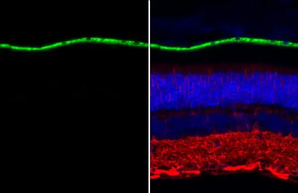
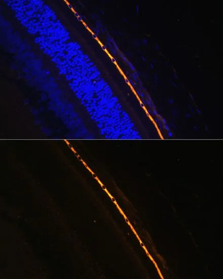
RPE65 antibody [N1C3] detects RPE65 protein at retinal pigment epithelium by immunohistochemical analysis. Sample: Paraffin-embedded mouse eye. Green: RPE65 stained by RPE65 antibody [N1C3] (GTX103472) diluted at 1:500. Red: beta Tubulin 3/ Tuj1, a neuronal marker, stained by beta Tubulin 3/ Tuj1 antibody [GT11710] (GTX631836) diluted at 1:500. Blue: Fluoroshield with DAPI (GTX30920). Antigen Retrieval: Citrate buffer, pH 6.0, 15 min
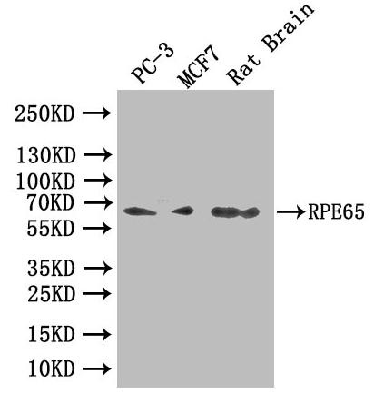
![Whole cell extract (30 μg) was separated by 7.5% SDS-PAGE, and the membrane was blotted with RPE65 antibody [N1C3] (GTX103472) diluted at 1:1000. The HRP-conjugated anti-rabbit IgG antibody (GTX213110-01) was used to detect the primary antibody.](https://www.genetex.com/upload/website/prouct_img/normal/GTX103472/GTX103472_44434_20211105_WB_25082900_161.webp "Whole cell extract (30 μg) was separated by 7.5% SDS-PAGE, and the membrane was blotted with RPE65 antibody [N1C3] (GTX103472) diluted at 1:1000. The HRP-conjugated anti-rabbit IgG antibody (GTX213110-01) was used to detect the primary antibody.")
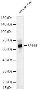
![Mouse tissue extract (50 μg) was separated by 7.5% SDS-PAGE, and the membrane was blotted with RPE65 antibody [N1C3] (GTX103472) diluted at 1:1000. The HRP-conjugated anti-rabbit IgG antibody (GTX213110-01) was used to detect the primary antibody.](https://www.genetex.com/upload/website/prouct_img/normal/GTX103472/GTX103472_44434_20210910_WB_M_eye_25082900_645.webp "Mouse tissue extract (50 μg) was separated by 7.5% SDS-PAGE, and the membrane was blotted with RPE65 antibody [N1C3] (GTX103472) diluted at 1:1000. The HRP-conjugated anti-rabbit IgG antibody (GTX213110-01) was used to detect the primary antibody.")
RPE65 antibody [N1C3] detects RPE65 protein at retinal pigment epithelium by immunohistochemical analysis. Sample: Paraffin-embedded mouse eye. Green: RPE65 stained by RPE65 antibody [N1C3] (GTX103472) diluted at 1:500. Red: beta Tubulin 3/ Tuj1, a neuronal marker, stained by beta Tubulin 3/ Tuj1 antibody [GT11710] (GTX631836) diluted at 1:500. Blue: Fluoroshield with DAPI (GTX30920). Antigen Retrieval: Citrate buffer, pH 6.0, 15 min
RPE65 antibody [N1C3]
GTX103472
ApplicationsImmunoFluorescence, Western Blot, ImmunoCytoChemistry, ImmunoHistoChemistry, ImmunoHistoChemistry Paraffin
Product group Antibodies
ReactivityHuman, Mouse
TargetRPE65
Overview
- SupplierGeneTex
- Product NameRPE65 antibody [N1C3]
- Delivery Days Customer9
- Application Supplier NoteWB: 1:500-1:3000. *Optimal dilutions/concentrations should be determined by the researcher.Not tested in other applications.
- ApplicationsImmunoFluorescence, Western Blot, ImmunoCytoChemistry, ImmunoHistoChemistry, ImmunoHistoChemistry Paraffin
- CertificationResearch Use Only
- ClonalityPolyclonal
- Concentration0.96 mg/ml
- ConjugateUnconjugated
- Gene ID6121
- Target nameRPE65
- Target descriptionretinoid isomerohydrolase RPE65
- Target synonymsBCO3, LCA2, RP20, mRPE65, p63, rd12, sRPE65, retinoid isomerohydrolase, BCO family, member 3, RBP-binding membrane protein, RPE65, retinoid isomerohydrolase, all-trans-retinyl-palmitate hydrolase, lutein isomerase, meso-zeaxanthin isomerase, retinal pigment epithelium specific protein 65, retinal pigment epithelium-specific 65 kDa protein, retinal pigment epithelium-specific protein 65kDa, retinitis pigmentosa 20 (autosomal recessive), retinol isomerase
- HostRabbit
- IsotypeIgG
- Protein IDQ16518
- Protein NameRetinoid isomerohydrolase
- Scientific DescriptionThis gene encodes a protein which is located in the retinal pigment epithelium and is involved in the production of 11-cis retinal and in visual pigment regeneration. There are two forms of this protein, a soluble form called sRPE65, and a palmitoylated, membrane-bound form known as mRPE65. mRPE65 serves as the palmitoyl donor for lecithin retinol acyl transferase (LRAT), the enzyme that catalyzes the vitamin A to all trans retinol step of the chromophore regeneration process. Both mRPE65 and sRPE65 also serve as regulatory proteins, with the ratio and concentrations of these molecules playing a role in the inhibition of 11-cis retinal synthesis. Mutations in this gene have been associated with Leber congenital amaurosis type 2 (LCA2) and retinitis pigmentosa. [provided by RefSeq]
- ReactivityHuman, Mouse
- Storage Instruction-20°C or -80°C,2°C to 8°C
- UNSPSC41116161
Datasheet
Related products
Product group Antibodies
Anti-RPE65 AntibodyA16350
ApplicationsImmunoFluorescence, Western Blot, ImmunoCytoChemistry
ReactivityHuman, Mouse, Rat
- SizePrice
Product group Antibodies
Anti-RPE65 Antibody144-09841
ApplicationsWestern Blot
ReactivityHuman, Mouse, Rat
TargetRPE65
- SizePrice
Product group Antibodies
RPE65 Recombinant Antibody, AbBy Fluor-488 ConjugatedBSM-61470R-BF488
ApplicationsImmunoFluorescence, Western Blot
ReactivityHuman, Mouse, Rat
TargetRPE65
- SizePrice
Product group Antibodies

Goat anti-RPE65EB08369
ApplicationsWestern Blot, ELISA
ReactivityCanine, Human, Rat
TargetRPE65
- SizePrice
Product group Antibodies
RPE65 AntibodyCSB-PA624103LA01HU
ApplicationsImmunoFluorescence, Western Blot, ELISA
ReactivityHuman, Rat
TargetRPE65
- SizePrice
Product group Antibodies
Rpe65 Polyclonal AntibodyCAC11395
ApplicationsImmunoFluorescence, Western Blot, ELISA
ReactivityRat
TargetRPE65
- SizePrice

Product group Antibodies
ApplicationsImmunoPrecipitation, Western Blot
ReactivityHuman, Mouse, Rat
TargetRPE65
- SizePrice
Product group Antibodies

RPE65 AntibodyLS-C411363
ApplicationsWestern Blot
ReactivityHuman, Mouse, Rat
TargetRPE65
- SizePrice
Product group Antibodies
RPE65 antibody, InternalGTX88833
ApplicationsWestern Blot
ReactivityHuman, Rat
TargetRPE65
- SizePrice
Product group Antibodies
RPE65 antibodyGTX64456
ApplicationsWestern Blot, ImmunoHistoChemistry, ImmunoHistoChemistry Paraffin
ReactivityHuman, Mouse, Rat
TargetRPE65
- SizePrice