

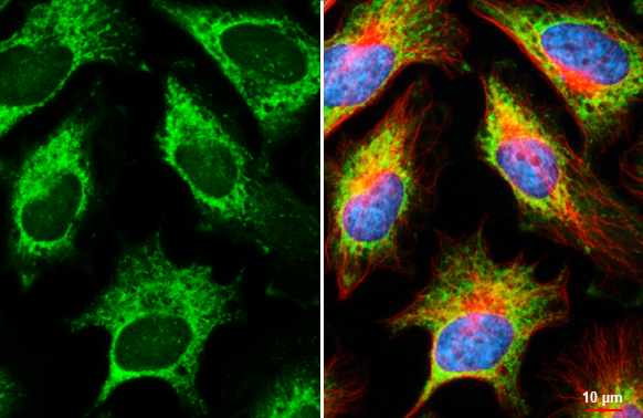
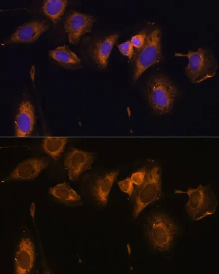
RPL32 antibody [HL2329] detects RPL32 protein at cytoplasm by immunofluorescent analysis. Sample: HeLa cells were fixed in ice-cold MeOH for 5 min. Green: RPL32 stained by RPL32 antibody [HL2329] (GTX638492) diluted at 1:500. Red: alpha Tubulin, a cytoskeleton marker, stained by alpha Tubulin antibody [GT114] (GTX628802) diluted at 1:1000. Blue: Fluoroshield with DAPI (GTX30920).
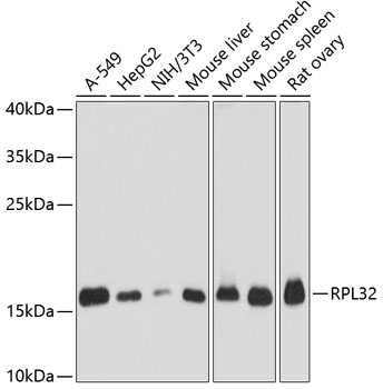
![Various tissue extracts (50 μg) were separated by 15% SDS-PAGE, and the membrane was blotted with RPL32 antibody [HL2329] (GTX638492) diluted at 1:1000. The HRP-conjugated anti-rabbit IgG antibody (GTX213110-01) was used to detect the primary antibody.](https://www.genetex.com/upload/website/prouct_img/normal/GTX638492/GTX638492_T-45005_20230609_WB_M_R_23061400_155.webp "Various tissue extracts (50 μg) were separated by 15% SDS-PAGE, and the membrane was blotted with RPL32 antibody [HL2329] (GTX638492) diluted at 1:1000. The HRP-conjugated anti-rabbit IgG antibody (GTX213110-01) was used to detect the primary antibody.")
![Various whole cell extracts (30 μg) were separated by 15% SDS-PAGE, and the membrane was blotted with RPL32 antibody [HL2329] (GTX638492) diluted at 1:1000. The HRP-conjugated anti-rabbit IgG antibody (GTX213110-01) was used to detect the primary antibody.](https://www.genetex.com/upload/website/prouct_img/normal/GTX638492/GTX638492_45103_20230714_WB_23071822_834.webp "Various whole cell extracts (30 μg) were separated by 15% SDS-PAGE, and the membrane was blotted with RPL32 antibody [HL2329] (GTX638492) diluted at 1:1000. The HRP-conjugated anti-rabbit IgG antibody (GTX213110-01) was used to detect the primary antibody.")
![Whole cell extract (30 μg) was separated by 15% SDS-PAGE, and the membrane was blotted with RPL32 antibody [HL2329] (GTX638492) diluted at 1:1000. The HRP-conjugated anti-rabbit IgG antibody (GTX213110-01) was used to detect the primary antibody.](https://www.genetex.com/upload/website/prouct_img/normal/GTX638492/GTX638492_45103_20230908_WB_D_23091319_140.webp "Whole cell extract (30 μg) was separated by 15% SDS-PAGE, and the membrane was blotted with RPL32 antibody [HL2329] (GTX638492) diluted at 1:1000. The HRP-conjugated anti-rabbit IgG antibody (GTX213110-01) was used to detect the primary antibody.")
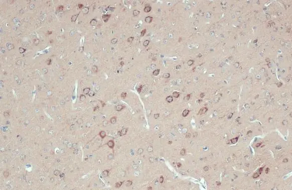
![RPL32 antibody [HL2329] detects RPL32 protein at cytoplasm by immunohistochemical analysis. Sample: Paraffin-embedded rat brain. RPL32 stained by RPL32 antibody [HL2329] (GTX638492) diluted at 1:100. Antigen Retrieval: Citrate buffer, pH 6.0, 15 min](https://www.genetex.com/upload/website/prouct_img/normal/GTX638492/GTX638492_T-45005_20230925_IHC-P_R_23100319_841.webp "RPL32 antibody [HL2329] detects RPL32 protein at cytoplasm by immunohistochemical analysis. Sample: Paraffin-embedded rat brain. RPL32 stained by RPL32 antibody [HL2329] (GTX638492) diluted at 1:100. Antigen Retrieval: Citrate buffer, pH 6.0, 15 min")
![RPL32 antibody [HL2329] detects RPL32 protein at cytoplasm by immunohistochemical analysis. Sample: Paraffin-embedded mouse brain. RPL32 stained by RPL32 antibody [HL2329] (GTX638492) diluted at 1:100. Antigen Retrieval: Citrate buffer, pH 6.0, 15 min](https://www.genetex.com/upload/website/prouct_img/normal/GTX638492/GTX638492_T-45005_20230925_IHC-P_M_23100319_569.webp "RPL32 antibody [HL2329] detects RPL32 protein at cytoplasm by immunohistochemical analysis. Sample: Paraffin-embedded mouse brain. RPL32 stained by RPL32 antibody [HL2329] (GTX638492) diluted at 1:100. Antigen Retrieval: Citrate buffer, pH 6.0, 15 min")
![Whole zebrafish extract (30 μg) was separated by 15% SDS-PAGE, and the membrane was blotted with RPL32 antibody [HL2329] (GTX638492) diluted at 1:3000. The HRP-conjugated anti-rabbit IgG antibody (GTX213110-01) was used to detect the primary antibody.](https://www.genetex.com/upload/website/prouct_img/normal/GTX638492/GTX638492_45103_20231006_WB_Z_23102401_539.webp "Whole zebrafish extract (30 μg) was separated by 15% SDS-PAGE, and the membrane was blotted with RPL32 antibody [HL2329] (GTX638492) diluted at 1:3000. The HRP-conjugated anti-rabbit IgG antibody (GTX213110-01) was used to detect the primary antibody.")
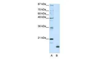
![Whole Japanese medaka extract (30 μg) was separated by 15% SDS-PAGE, and the membrane was blotted with RPL32 antibody [HL2329] (GTX638492) diluted at 1:1000. The HRP-conjugated anti-rabbit IgG antibody (GTX213110-01) was used to detect the primary antibody.](https://www.genetex.com/upload/website/prouct_img/normal/GTX638492/GTX638492_45103_20250815_WB_medaka_25082121_623.webp "Whole Japanese medaka extract (30 μg) was separated by 15% SDS-PAGE, and the membrane was blotted with RPL32 antibody [HL2329] (GTX638492) diluted at 1:1000. The HRP-conjugated anti-rabbit IgG antibody (GTX213110-01) was used to detect the primary antibody.")
RPL32 antibody [HL2329] detects RPL32 protein at cytoplasm by immunofluorescent analysis. Sample: HeLa cells were fixed in ice-cold MeOH for 5 min. Green: RPL32 stained by RPL32 antibody [HL2329] (GTX638492) diluted at 1:500. Red: alpha Tubulin, a cytoskeleton marker, stained by alpha Tubulin antibody [GT114] (GTX628802) diluted at 1:1000. Blue: Fluoroshield with DAPI (GTX30920).
RPL32 antibody [HL2329]
GTX638492
ApplicationsImmunoFluorescence, Western Blot, ImmunoCytoChemistry, ImmunoHistoChemistry, ImmunoHistoChemistry Paraffin
Product group Antibodies
ReactivityCanine, Human, Mouse, Rat, Zebra Fish
TargetRPL32
Overview
- SupplierGeneTex
- Product NameRPL32 antibody [HL2329]
- Delivery Days Customer9
- Application Supplier NoteWB: 1:500-1:10000. ICC/IF: 1:100-1:1000. *Optimal dilutions/concentrations should be determined by the researcher.Not tested in other applications.
- ApplicationsImmunoFluorescence, Western Blot, ImmunoCytoChemistry, ImmunoHistoChemistry, ImmunoHistoChemistry Paraffin
- CertificationResearch Use Only
- ClonalityMonoclonal
- Clone IDHL2329
- Concentration1 mg/ml
- ConjugateUnconjugated
- Gene ID6161
- Target nameRPL32
- Target descriptionribosomal protein L32
- Target synonymsL32, PP9932, eL32, large ribosomal subunit protein eL32, 60S ribosomal protein L32
- HostRabbit
- IsotypeIgG
- Protein IDP62910
- Protein NameLarge ribosomal subunit protein eL32
- Scientific DescriptionRibosomes, the organelles that catalyze protein synthesis, consist of a small 40S subunit and a large 60S subunit. Together these subunits are composed of 4 RNA species and approximately 80 structurally distinct proteins. This gene encodes a ribosomal protein that is a component of the 60S subunit. The protein belongs to the L32E family of ribosomal proteins. It is located in the cytoplasm. Although some studies have mapped this gene to 3q13.3-q21, it is believed to map to 3p25-p24. As is typical for genes encoding ribosomal proteins, there are multiple processed pseudogenes of this gene dispersed through the genome. Alternatively spliced transcript variants encoding the same protein have been observed for this gene. [provided by RefSeq, Jul 2008]
- ReactivityCanine, Human, Mouse, Rat, Zebra Fish
- Storage Instruction-20°C or -80°C,2°C to 8°C
- UNSPSC41116161
Datasheet
Related products
Product group Antibodies
RPL32 AntibodyCSB-PA020237GA01HU
ApplicationsWestern Blot, ELISA
ReactivityHuman, Mouse, Rat
TargetRPL32
- SizePrice

Product group Antibodies
Anti-RPL32 Antibody Picoband(r)A06487-1-CARRIER-FREE
ApplicationsFlow Cytometry, ImmunoFluorescence, Western Blot, ELISA, ImmunoCytoChemistry, ImmunoHistoChemistry
ReactivityHuman, Mouse, Rat
TargetRPL32
- SizePrice
Product group Antibodies
Anti-RPL32 AntibodyA88458
ApplicationsImmunoFluorescence, Western Blot, ImmunoCytoChemistry, ImmunoHistoChemistry
ReactivityHuman, Mouse, Rat
- SizePrice
Product group Antibodies

RPL32 / Ribosomal Protein L32 AntibodyLS-C748066
ApplicationsWestern Blot
ReactivityHuman, Mouse, Rat
TargetRPL32
- SizePrice

Product group Antibodies
Anti-RPL32 AntibodyHPA047501
ApplicationsWestern Blot, ImmunoHistoChemistry
ReactivityHuman
TargetRPL32
- SizePrice
![Various tissue extracts (50 μg) were separated by 15% SDS-PAGE, and the membrane was blotted with RPL32 antibody [HL2331] (GTX638494) diluted at 1:1000. The HRP-conjugated anti-rabbit IgG antibody (GTX213110-01) was used to detect the primary antibody.](https://www.genetex.com/upload/website/prouct_img/normal/GTX638494/GTX638494_T-45005_20230421_WB_M_R_23042500_392.webp)
Product group Antibodies
RPL32 antibody [HL2331]GTX638494
ApplicationsImmunoFluorescence, Western Blot, ImmunoCytoChemistry, ImmunoHistoChemistry, ImmunoHistoChemistry Paraffin
ReactivityCanine, Human, Mouse, Rat, Zebra Fish
TargetRPL32
- SizePrice
Product group Antibodies
RPL32 antibodyGTX66149
ApplicationsImmunoFluorescence, Western Blot, ImmunoCytoChemistry, ImmunoHistoChemistry, ImmunoHistoChemistry Paraffin
ReactivityHuman, Mouse, Rat
TargetRPL32
- SizePrice
Product group Antibodies
RPL32 antibodyGTX130214
ApplicationsImmunoFluorescence, Western Blot, ImmunoCytoChemistry, ImmunoHistoChemistry, ImmunoHistoChemistry Paraffin
ReactivityHuman, Mouse, Rat
TargetRPL32
- SizePrice
Product group Antibodies
RPL32 antibody, N-termGTX47428
ApplicationsWestern Blot
ReactivityHuman
TargetRPL32
- SizePrice