

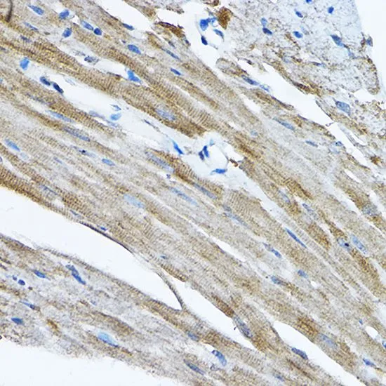
IHC-P analysis of mouse heart tissue using GTX32847 RPL36 antibody. Dilution : 1:200


IHC-P analysis of mouse heart tissue using GTX32847 RPL36 antibody. Dilution : 1:200
RPL36 antibody
GTX32847
ApplicationsImmunoFluorescence, Western Blot, ImmunoCytoChemistry, ImmunoHistoChemistry, ImmunoHistoChemistry Paraffin
Product group Antibodies
ReactivityHuman, Mouse, Rat
TargetRPL36
Overview
- SupplierGeneTex
- Product NameRPL36 antibody
- Delivery Days Customer9
- Application Supplier NoteWB: 1:500 - 1:2000. ICC/IF: 1:50 - 1:200. IHC-P: 1:50 - 1:200. *Optimal dilutions/concentrations should be determined by the researcher.Not tested in other applications.
- ApplicationsImmunoFluorescence, Western Blot, ImmunoCytoChemistry, ImmunoHistoChemistry, ImmunoHistoChemistry Paraffin
- CertificationResearch Use Only
- ClonalityPolyclonal
- ConjugateUnconjugated
- Gene ID25873
- Target nameRPL36
- Target descriptionribosomal protein L36
- Target synonymsL36, eL36, large ribosomal subunit protein eL36, 60S ribosomal protein L36
- HostRabbit
- IsotypeIgG
- Protein IDQ9Y3U8
- Protein NameLarge ribosomal subunit protein eL36
- Scientific DescriptionRibosomes, the organelles that catalyze protein synthesis, consist of a small 40S subunit and a large 60S subunit. Together these subunits are composed of 4 RNA species and approximately 80 structurally distinct proteins. This gene encodes a ribosomal protein that is a component of the 60S subunit. The protein belongs to the L36E family of ribosomal proteins. It is located in the cytoplasm. Transcript variants derived from alternative splicing exist; they encode the same protein. As is typical for genes encoding ribosomal proteins, there are multiple processed pseudogenes of this gene dispersed through the genome. [provided by RefSeq, Jul 2008]
- ReactivityHuman, Mouse, Rat
- Storage Instruction-20°C or -80°C,2°C to 8°C
- UNSPSC12352203
Datasheet
Related products

Product group Antibodies
Anti-RPL36 Antibody Picoband(r)A08922-3-CARRIER-FREE
ApplicationsFlow Cytometry, Western Blot, ImmunoHistoChemistry
ReactivityHuman, Monkey, Mouse, Rat
TargetRPL36
- SizePrice
Product group Antibodies
Anti-RPL36 Antibody144-07793
ApplicationsImmunoFluorescence, Western Blot, ImmunoHistoChemistry
ReactivityHuman, Mouse, Rat
TargetRPL36
- SizePrice
Product group Antibodies
RPL36 AntibodyCSB-PA003991
ApplicationsImmunoFluorescence, Western Blot, ELISA, ImmunoHistoChemistry
ReactivityHuman, Mouse, Rat
TargetRPL36
- SizePrice
Product group Antibodies
Anti-RPL36 AntibodyA95794
ApplicationsWestern Blot, ELISA, ImmunoHistoChemistry
ReactivityHuman, Mouse, Rat
- SizePrice
Product group Antibodies
Anti-RPL36 AntibodyHPA047153
ApplicationsWestern Blot, ImmunoHistoChemistry
ReactivityHuman
TargetRPL36
- SizePrice
Product group Antibodies

RPL36 / Ribosomal Protein L36 AntibodyLS-C409342
ApplicationsWestern Blot, ImmunoHistoChemistry
ReactivityHuman, Mouse, Rat
TargetRPL36
- SizePrice