

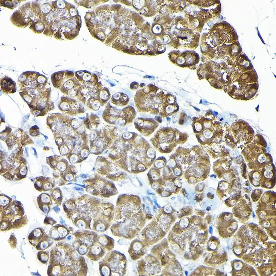
IHC-P analysis of rat pancreas tissue using GTX32849 RPS14 antibody. Dilution : 1:200


IHC-P analysis of rat pancreas tissue using GTX32849 RPS14 antibody. Dilution : 1:200
RPS14 antibody
GTX32849
ApplicationsImmunoFluorescence, Western Blot, ImmunoCytoChemistry, ImmunoHistoChemistry, ImmunoHistoChemistry Paraffin
Product group Antibodies
ReactivityHuman, Mouse, Rat
TargetRPS14
Overview
- SupplierGeneTex
- Product NameRPS14 antibody
- Delivery Days Customer9
- Application Supplier NoteWB: 1:500 - 1:2000. ICC/IF: 1:50 - 1:200. IHC-P: 1:50 - 1:200. *Optimal dilutions/concentrations should be determined by the researcher.Not tested in other applications.
- ApplicationsImmunoFluorescence, Western Blot, ImmunoCytoChemistry, ImmunoHistoChemistry, ImmunoHistoChemistry Paraffin
- CertificationResearch Use Only
- ClonalityPolyclonal
- ConjugateUnconjugated
- Gene ID6208
- Target nameRPS14
- Target descriptionribosomal protein S14
- Target synonymsEMTB, S14, uS11, small ribosomal subunit protein uS11, 40S ribosomal protein S14, emetine resistance
- HostRabbit
- IsotypeIgG
- Protein IDP62263
- Protein NameSmall ribosomal subunit protein uS11
- Scientific DescriptionRibosomes, the organelles that catalyze protein synthesis, consist of a small 40S subunit and a large 60S subunit. Together these subunits are composed of 4 RNA species and approximately 80 structurally distinct proteins. This gene encodes a ribosomal protein that is a component of the 40S subunit. The protein belongs to the S11P family of ribosomal proteins. It is located in the cytoplasm. Transcript variants utilizing alternative transcription initiation sites have been described in the literature. As is typical for genes encoding ribosomal proteins, there are multiple processed pseudogenes of this gene dispersed through the genome. In Chinese hamster ovary cells, mutations in this gene can lead to resistance to emetine, a protein synthesis inhibitor. Multiple alternatively spliced transcript variants encoding the same protein have been found for this gene. [provided by RefSeq, Jul 2008]
- ReactivityHuman, Mouse, Rat
- Storage Instruction-20°C or -80°C,2°C to 8°C
- UNSPSC41116161
Datasheet
Related products
Product group Antibodies
Anti-RPS14 AntibodyA31556
ApplicationsImmunoFluorescence, Western Blot, ImmunoHistoChemistry
ReactivityHuman, Mouse, Rat
- SizePrice

Product group Antibodies
Anti-RPS14 Antibody Picoband(r)A03696-1-CARRIER-FREE
ApplicationsFlow Cytometry, Western Blot, ELISA, ImmunoHistoChemistry
ReactivityHuman, Mouse, Rat
TargetRPS14
- SizePrice
Product group Antibodies
Anti-RPS14 Antibody144-06727
ApplicationsImmunoFluorescence, Western Blot, ImmunoHistoChemistry
ReactivityHuman, Mouse, Rat
TargetRPS14
- SizePrice
Product group Antibodies

RPS14 / Ribosomal Protein S14 AntibodyLS-C830886
ApplicationsWestern Blot, ELISA, ImmunoHistoChemistry
ReactivityHuman, Mouse, Rat
TargetRPS14
- SizePrice
Product group Antibodies
RPS14 AntibodyCSB-PA04255A0RB
ApplicationsELISA
ReactivityHuman
TargetRPS14
- SizePrice

Product group Antibodies
Anti-RPS14 AntibodyHPA018504
ApplicationsWestern Blot, ImmunoCytoChemistry, ImmunoHistoChemistry
ReactivityHuman, Mouse
TargetRPS14
- SizePrice
Product group Antibodies
Anti-RPS14 AntibodyCAB6727
ApplicationsImmunoFluorescence, Western Blot, ELISA, ImmunoCytoChemistry, ImmunoHistoChemistry, ImmunoHistoChemistry Paraffin
ReactivityHuman
TargetRPS14
- SizePrice