

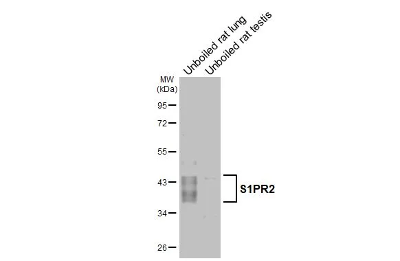
Unboiled various tissue extracts (50 μg) were separated by 10% SDS-PAGE, and the membrane was blotted with S1PR2 antibody [HL3765] (GTX641964) diluted at 1:1000. The HRP-conjugated anti-rabbit IgG antibody (GTX213110-01) was used to detect the primary antibody, and the signal was developed with Trident ECL plus-Enhanced.
![Boiled and unboiled HCT116 whole cell and membrane extracts (30 μg) were separated by 10% SDS-PAGE, and the membrane was blotted with S1PR2 antibody [HL3765] (GTX641964) diluted at 1:1000. The HRP-conjugated anti-rabbit IgG antibody (GTX213110-01) was used to detect the primary antibody.(WCE: whole cell extract; ME: membrane extract)](https://www.genetex.com/upload/website/prouct_img/normal/GTX641964/GTX641964_T-45670_20250502_WB_Fraction_ub_25050623_723.webp "Boiled and unboiled HCT116 whole cell and membrane extracts (30 μg) were separated by 10% SDS-PAGE, and the membrane was blotted with S1PR2 antibody [HL3765] (GTX641964) diluted at 1:1000. The HRP-conjugated anti-rabbit IgG antibody (GTX213110-01) was used to detect the primary antibody.(WCE: whole cell extract; ME: membrane extract)")
![Unboiled various tissue extracts (50 μg) were separated by 10% SDS-PAGE, and the membrane was blotted with S1PR2 antibody [HL3765] (GTX641964) diluted at 1:1000. The HRP-conjugated anti-rabbit IgG antibody (GTX213110-01) was used to detect the primary antibody. Corresponding RNA expression data are based on NCBI database.](https://www.genetex.com/upload/website/prouct_img/normal/GTX641964/GTX641964_T-45670_20250502_WB_M_tissue_RPKM_watermark_25050623_454.webp "Unboiled various tissue extracts (50 μg) were separated by 10% SDS-PAGE, and the membrane was blotted with S1PR2 antibody [HL3765] (GTX641964) diluted at 1:1000. The HRP-conjugated anti-rabbit IgG antibody (GTX213110-01) was used to detect the primary antibody. Corresponding RNA expression data are based on NCBI database.")
![Unboiled Raji whole cell and membrane extracts (30 μg) were separated by 10% SDS-PAGE, and the membrane was blotted with S1PR2 antibody [HL3765] (GTX641964) diluted at 1:1000. The HRP-conjugated anti-rabbit IgG antibody (GTX213110-01) was used to detect the primary antibody. (WCE: whole cell extract; ME: membrane extract)](https://www.genetex.com/upload/website/prouct_img/normal/GTX641964/GTX641964_T-45670_20250502_WB_Fraction_25050623_529.webp "Unboiled Raji whole cell and membrane extracts (30 μg) were separated by 10% SDS-PAGE, and the membrane was blotted with S1PR2 antibody [HL3765] (GTX641964) diluted at 1:1000. The HRP-conjugated anti-rabbit IgG antibody (GTX213110-01) was used to detect the primary antibody. (WCE: whole cell extract; ME: membrane extract)")
![Unboiled various tissue extracts (30 μg) were separated by 10% SDS-PAGE, and the membrane was blotted with S1PR2 antibody [HL3765] (GTX641964) diluted at 1:2400. The HRP-conjugated anti-rabbit IgG antibody (GTX213110-01) was used to detect the primary antibody. Corresponding RNA expression data are based on NCBI database.](https://www.genetex.com/upload/website/prouct_img/normal/GTX641964/GTX641964_T-45670_20250516_WB_M_tissue_RPKM_watermark_25052202_943.webp "Unboiled various tissue extracts (30 μg) were separated by 10% SDS-PAGE, and the membrane was blotted with S1PR2 antibody [HL3765] (GTX641964) diluted at 1:2400. The HRP-conjugated anti-rabbit IgG antibody (GTX213110-01) was used to detect the primary antibody. Corresponding RNA expression data are based on NCBI database.")
 detects S1PR2 protein by flow cytometry analysis. Sample: Non-transfected and transfected 293T cells were fixed in 4% paraformaldehyde at 4oC for 15 min and washed with 0.1% saponin buffer. The cells were following stained with S1PR2 antibody [HL3765](GTX641964) diluted at 1:50 or a Rabbit IgG isotype control (GTX35035) at 4oC for 1 hour.](https://www.genetex.com/upload/website/prouct_img/normal/GTX641964/GTX641964_45810_20251017_FCM_25112100_585.webp "S1PR2 antibody [HL3765](GTX641964) detects S1PR2 protein by flow cytometry analysis. Sample: Non-transfected and transfected 293T cells were fixed in 4% paraformaldehyde at 4oC for 15 min and washed with 0.1% saponin buffer. The cells were following stained with S1PR2 antibody [HL3765](GTX641964) diluted at 1:50 or a Rabbit IgG isotype control (GTX35035) at 4oC for 1 hour.")
Unboiled various tissue extracts (50 μg) were separated by 10% SDS-PAGE, and the membrane was blotted with S1PR2 antibody [HL3765] (GTX641964) diluted at 1:1000. The HRP-conjugated anti-rabbit IgG antibody (GTX213110-01) was used to detect the primary antibody, and the signal was developed with Trident ECL plus-Enhanced.
S1PR2 antibody [HL3765]
GTX641964
ApplicationsFlow Cytometry, Western Blot
Product group Antibodies
ReactivityHuman, Mouse, Rat
TargetS1PR2
Overview
- SupplierGeneTex
- Product NameS1PR2 antibody [HL3765]
- Delivery Days Customer9
- Application Supplier NoteWB: 1:500-1:3000. *Optimal dilutions/concentrations should be determined by the researcher.Not tested in other applications.
- ApplicationsFlow Cytometry, Western Blot
- CertificationResearch Use Only
- ClonalityMonoclonal
- Clone IDHL3765
- Concentration1 mg/ml
- ConjugateUnconjugated
- Gene ID9294
- Target nameS1PR2
- Target descriptionsphingosine-1-phosphate receptor 2
- Target synonymsAGR16, DFNB68, EDG-5, EDG5, Gpcr13, H218, LPB2, S1P2, sphingosine 1-phosphate receptor 2, CTD-2369P2.2, S1P receptor 2, S1P receptor EDG5, S1P receptor Edg-5, deafness, autosomal recessive 68, endothelial differentiation G-protein coupled receptor 5, endothelial differentiation, sphingolipid G-protein-coupled receptor, 5, sphingosine 1-phosphate receptor Edg-5
- HostRabbit
- IsotypeIgG
- Protein IDO95136
- Protein NameSphingosine 1-phosphate receptor 2
- Scientific DescriptionThis gene encodes a member of the G protein-coupled receptors, as well as the EDG family of proteins. The encoded protein is a receptor for sphingosine 1-phosphate, which participates in cell proliferation, survival, and transcriptional activation. Defects in this gene have been associated with congenital profound deafness. [provided by RefSeq, Mar 2016]
- ReactivityHuman, Mouse, Rat
- Storage Instruction-20°C or -80°C,2°C to 8°C
- UNSPSC41116161
Related products

Product group Antibodies
Anti-EDG5 AntibodyA98186
ApplicationsWestern Blot, ELISA
ReactivityHuman, Mouse, Rat
- SizePrice
Product group Antibodies
S1PR2 AntibodyCSB-PA002249
ApplicationsImmunoFluorescence, Western Blot, ELISA, ImmunoHistoChemistry
ReactivityHuman, Mouse, Rat
TargetS1PR2
- SizePrice
Product group Antibodies

Goat anti-S1PR2EB10252
ApplicationsWestern Blot, ELISA
ReactivityCanine, Human, Mouse, Rat
TargetS1PR2
- SizePrice
Product group Antibodies
S1PR2 Polyclonal AntibodyCAC15061
ApplicationsImmunoFluorescence, Western Blot, ELISA, ImmunoHistoChemistry
ReactivityMouse
TargetS1PR2
- SizePrice
Product group Antibodies

ApplicationsImmunoFluorescence, Western Blot, ImmunoCytoChemistry, ImmunoHistoChemistry
ReactivityHuman, Mouse, Rat
TargetS1PR2
- SizePrice
Product group Antibodies
S1PR2 antibodyGTX87713
ApplicationsWestern Blot
ReactivityHuman
TargetS1PR2
- SizePrice