

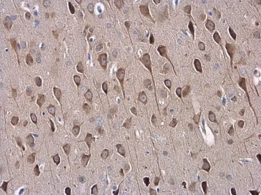
SAP102 antibody [C1C3] detects SAP102 protein at cytoplasm in mouse brain by immunohistochemical analysis. Sample: Paraffin-embedded mouse brain. SAP102 antibody [C1C3] (GTX110289) diluted at 1:500.
Antigen Retrieval: Citrate buffer, pH 6.0, 15 min
![Various tissue extracts (50 μg) were separated by 7.5% SDS-PAGE, and the membrane was blotted with SAP102 antibody [C1C3] (GTX110289) diluted at 1:1000.](https://www.genetex.com/upload/website/prouct_img/normal/GTX110289/GTX110289_40037_20160922_WB_M_R_w_23060500_461.webp "Various tissue extracts (50 μg) were separated by 7.5% SDS-PAGE, and the membrane was blotted with SAP102 antibody [C1C3] (GTX110289) diluted at 1:1000.")
 A: Molt-4 (GTX27912) 7.5% SDS PAGE GTX110298 diluted at 1:1000")
![SAP102 antibody [C1C3] detects SAP102 protein expression by immunohistochemical analysis. Sample: Frozen sectioned E13.5 Rat brain. Green: SAP102 antibody [C1C3] stained by SAP102 antibody (GTX110289) diluted at 1:250. Red: beta Tubulin 3/ TUJ1, a mature neuron marker, stained by beta Tubulin 3/ TUJ1 antibody [GT11710] (GTX631836) diluted at 1:500. Blue: Fluoroshield with DAPI (GTX30920).](https://www.genetex.com/upload/website/prouct_img/normal/GTX110289/GTX110289_40037_20161012_IHC-Fr_R_w_23060500_509.webp "SAP102 antibody [C1C3] detects SAP102 protein expression by immunohistochemical analysis. Sample: Frozen sectioned E13.5 Rat brain. Green: SAP102 antibody [C1C3] stained by SAP102 antibody (GTX110289) diluted at 1:250. Red: beta Tubulin 3/ TUJ1, a mature neuron marker, stained by beta Tubulin 3/ TUJ1 antibody [GT11710] (GTX631836) diluted at 1:500. Blue: Fluoroshield with DAPI (GTX30920).")
![SAP102 antibody [C1C3] detects SAP102 protein at cytoplasm by immunofluorescent analysis. Sample: HeLa cells were fixed in 4% paraformaldehyde at RT for 15 min. Green: SAP102 protein stained by SAP102 antibody [C1C3] (GTX110289) diluted at 1:500. Red: alpha Tubulin, a cytoskeleton marker, stained by alpha Tubulin antibody [GT114] (GTX628802) diluted at 1:500. Blue: Hoechst 33342 staining.](https://www.genetex.com/upload/website/prouct_img/normal/GTX110289/GTX110289_40037_20150410_IFA_w_23060500_689.webp "SAP102 antibody [C1C3] detects SAP102 protein at cytoplasm by immunofluorescent analysis. Sample: HeLa cells were fixed in 4% paraformaldehyde at RT for 15 min. Green: SAP102 protein stained by SAP102 antibody [C1C3] (GTX110289) diluted at 1:500. Red: alpha Tubulin, a cytoskeleton marker, stained by alpha Tubulin antibody [GT114] (GTX628802) diluted at 1:500. Blue: Hoechst 33342 staining.")
 antibody at 1:200 dilution.")
![SAP102 antibody [C1C3] detects SAP102 protein at cell body and synaptic vesicles by immunofluorescent analysis. Sample: DIV9 rat E18 primary cortical neurons were fixed in 4% paraformaldehyde at RT for 15 min. Green: SAP102 protein stained by SAP102 antibody [C1C3] (GTX110289) diluted at 1:500. Red: beta Tubulin 3/ Tuj1, a neuron cell marker, stained by beta Tubulin 3/ Tuj1 antibody [GT11710] (GTX631836) diluted at 1:500. Blue: Fluoroshield with DAPI (GTX30920).](https://www.genetex.com/upload/website/prouct_img/normal/GTX110289/GTX110289_40037_20170727_IFA_R_w_23060500_691.webp "SAP102 antibody [C1C3] detects SAP102 protein at cell body and synaptic vesicles by immunofluorescent analysis. Sample: DIV9 rat E18 primary cortical neurons were fixed in 4% paraformaldehyde at RT for 15 min. Green: SAP102 protein stained by SAP102 antibody [C1C3] (GTX110289) diluted at 1:500. Red: beta Tubulin 3/ Tuj1, a neuron cell marker, stained by beta Tubulin 3/ Tuj1 antibody [GT11710] (GTX631836) diluted at 1:500. Blue: Fluoroshield with DAPI (GTX30920).")
 A: Mouse brain 7.5% SDS PAGE GTX110289 diluted at 1:1000")
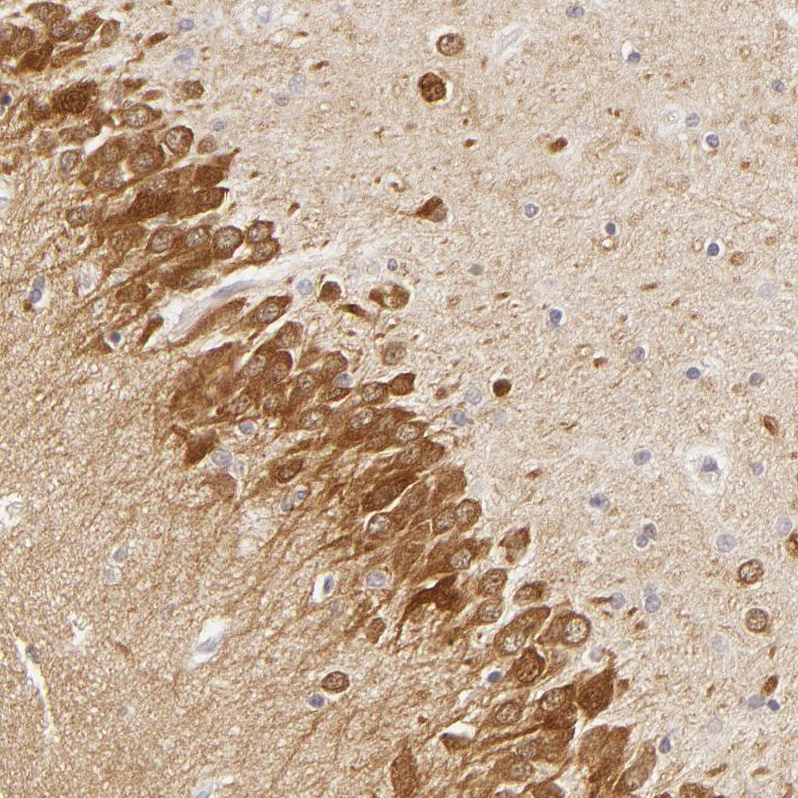
![SAP102 antibody [C1C3] detects SAP102 protein at cytoplasm in mouse brain by immunohistochemical analysis. Sample: Paraffin-embedded mouse brain. SAP102 antibody [C1C3] (GTX110289) diluted at 1:500.
Antigen Retrieval: Citrate buffer, pH 6.0, 15 min](https://www.genetex.com/upload/website/prouct_img/normal/GTX110289/GTX110289_40037_20160301_IHC-P_M_2_w_23060500_436.webp "SAP102 antibody [C1C3] detects SAP102 protein at cytoplasm in mouse brain by immunohistochemical analysis. Sample: Paraffin-embedded mouse brain. SAP102 antibody [C1C3] (GTX110289) diluted at 1:500.
Antigen Retrieval: Citrate buffer, pH 6.0, 15 min")
SAP102 antibody [C1C3] detects SAP102 protein at cytoplasm in mouse brain by immunohistochemical analysis. Sample: Paraffin-embedded mouse brain. SAP102 antibody [C1C3] (GTX110289) diluted at 1:500.
Antigen Retrieval: Citrate buffer, pH 6.0, 15 min
SAP102 antibody [C1C3]
GTX110289
ApplicationsImmunoFluorescence, Western Blot, ImmunoCytoChemistry, ImmunoHistoChemistry, ImmunoHistoChemistry Frozen, ImmunoHistoChemistry Paraffin
Product group Antibodies
ReactivityHuman, Mouse, Porcine, Rat
TargetDLG3
Overview
- SupplierGeneTex
- Product NameSAP102 antibody [C1C3]
- Delivery Days Customer9
- Application Supplier NoteWB: 1:500-1:3000. ICC/IF: 1:100-1:1000. IHC-P: 1:100-1:1000. IHC-Fr: 1:100-1:1000. *Optimal dilutions/concentrations should be determined by the researcher.Not tested in other applications.
- ApplicationsImmunoFluorescence, Western Blot, ImmunoCytoChemistry, ImmunoHistoChemistry, ImmunoHistoChemistry Frozen, ImmunoHistoChemistry Paraffin
- CertificationResearch Use Only
- ClonalityPolyclonal
- Concentration1 mg/ml
- ConjugateUnconjugated
- Gene ID1741
- Target nameDLG3
- Target descriptiondiscs large MAGUK scaffold protein 3
- Target synonymsMRX, MRX90, NEDLG, PPP1R82, SAP102, XLID90, XLMR, disks large homolog 3, discs, large homolog 3, neuroendocrine-DLG, protein phosphatase 1, regulatory subunit 82, synapse-associated protein 102
- HostRabbit
- IsotypeIgG
- Protein IDQ92796
- Protein NameDisks large homolog 3
- Scientific DescriptionThis gene encodes a member of the membrane-associated guanylate kinase protein family. The encoded protein may play a role in clustering of NMDA receptors at excitatory synapses. It may also negatively regulate cell proliferation through interaction with the C-terminal region of the adenomatosis polyposis coli tumor suppressor protein. Mutations in this gene have been associated with X-linked mental retardation. Alternatively spliced transcript variants have been described. [provided by RefSeq]
- ReactivityHuman, Mouse, Porcine, Rat
- Storage Instruction-20°C or -80°C,2°C to 8°C
- UNSPSC41116161
Datasheet
Related products
Product group Antibodies
DLG3 AntibodyCSB-PA006937GA01HU
ApplicationsWestern Blot, ELISA
ReactivityHuman, Mouse, Rat
TargetDLG3
- SizePrice

Product group Antibodies
Anti-SAP102 AntibodyA93210
ApplicationsWestern Blot, ImmunoHistoChemistry
ReactivityHuman, Mouse, Rat
- SizePrice
Product group Antibodies
Anti-DLG3 AntibodyHPA001733
ApplicationsWestern Blot, ImmunoHistoChemistry
ReactivityHuman
TargetDLG3
- SizePrice

Product group Antibodies
Anti-SAP102/DLG3 Antibody Picoband(r)A04152-1-CARRIER-FREE
ApplicationsWestern Blot, ImmunoHistoChemistry
ReactivityHuman, Mouse, Rat
TargetDLG3
- SizePrice
Product group Antibodies

DLG3 / SAP102 Antibody (aa627-802)LS-C373505
ApplicationsWestern Blot
ReactivityHuman, Porcine, Rat
TargetDLG3
- SizePrice
Product group Antibodies
ApplicationsImmunoPrecipitation, Western Blot, ImmunoCytoChemistry, ImmunoHistoChemistry
ReactivityMouse, Porcine, Rat
TargetDLG3
- SizePrice