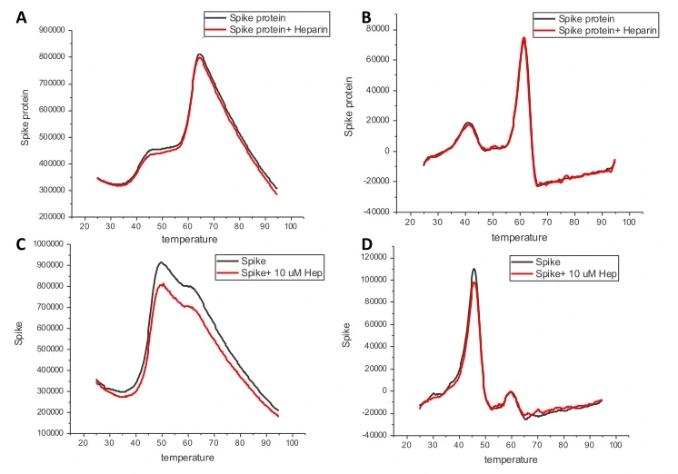
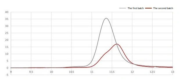
Differential scanning fluorimetry (DFS) analysis the stability and the heparin binding ability. DSF of proteins were performed in the absence or presence of heparin (10 μM). The scanning fluorimetry data of the two batches shows the protein is both stable and functional, with heparin binding activity. A and C : Melting curve of spike protein. B and D : First derivative of the melting curves of spike to show its melting temperature as peak. A and B : the first batch of spike protein (PBS) C and D : the second batch of spike protein (PBS, 20% glycerol).
SARS-CoV-2 (COVID-19) Spike (D614G Mutant)(ECD) protein, His tag (active)
GTX02575-PRO
ApplicationsFunctional Assay, ELISA
Product group Proteins / Signaling Molecules
Overview
- SupplierGeneTex
- Product NameSARS-CoV-2 (COVID-19) Spike (D614G Mutant)(ECD) protein, His tag (active)
- Delivery Days Customer9
- ApplicationsFunctional Assay, ELISA
- CertificationResearch Use Only
- ConjugateUnconjugated
- Gene ID43740568
- Target nameS
- Target descriptionsurface glycoprotein
- Target synonymsGU280_gp02, spike glycoprotein, surface glycoprotein
- Storage Instruction-20°C
- UNSPSC41116100
- SpeciesVirus

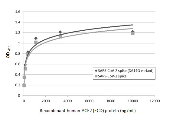
![Sandwich ELISA detection of recombinant SARS-CoV-2 (COVID-19) Spike (D614G variant) protein, His tag (active) (GTX02575-pro), and SARS-CoV-2 spike (trimer) protein using SARS-CoV-2 (COVID-19) Spike RBD antibody [HL1014] (GTX635807) as capture antibody at concentration of 5 μg/mL and SARS-CoV-2 (COVID-19) Spike RBD antibody [HL1003] (HRP) (GTX635792-01) as detection antibody at concentration of 1 μg/mL. Sandwich ELISA detection of recombinant SARS-CoV-2 (COVID-19) Spike (D614G variant) protein, His tag (active) (GTX02575-pro), and SARS-CoV-2 spike (trimer) protein using SARS-CoV-2 (COVID-19) Spike RBD antibody [HL1014] (GTX635807) as capture antibody at concentration of 5 μg/mL and SARS-CoV-2 (COVID-19) Spike RBD antibody [HL1003] (HRP) (GTX635792-01) as detection antibody at concentration of 1 μg/mL.](https://www.genetex.com/upload/website/prouct_img/normal/GTX02575-pro/GTX02575-pro_822005406_20210305_ELISA_PAIR_w_23053122_679.webp)