

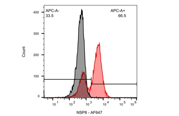
SARS-CoV / SARS-CoV-2 (COVID-19) NSP8 antibody [5A10] (GTX632696) detects SARS-CoV-2 (COVID-19) NSP8 by flow cytometry analysis. Sample: Vero E6 cells infected with SARS-CoV-2. Black: Uninfected Vero E6 cells was used as a control. Red: SARS-CoV / SARS-CoV-2 (COVID-19) NSP8 antibody [5A10] (GTX632696) dilution: 1:100.
![SARS-CoV / SARS-CoV-2 (COVID-19) NSP8 antibody [5A10] detects SARS-CoV / SARS-CoV-2 (COVID-19) NSP8 protein by immunofluorescent analysis. Sample: Mock and transfected 293T cells were fixed in 4% paraformaldehyde at RT for 15 min. Green: SARS-CoV / SARS-CoV-2 (COVID-19) NSP8 stained by SARS-CoV / SARS-CoV-2 (COVID-19) NSP8 antibody [5A10] (GTX632696) diluted at 1:1000. Blue: Fluoroshield with DAPI (GTX30920).](https://www.genetex.com/upload/website/prouct_img/normal/GTX632696/GTX632696_43969_20200708_ICC_IF_SARScoronavirus_B_w_23061202_409.webp "SARS-CoV / SARS-CoV-2 (COVID-19) NSP8 antibody [5A10] detects SARS-CoV / SARS-CoV-2 (COVID-19) NSP8 protein by immunofluorescent analysis. Sample: Mock and transfected 293T cells were fixed in 4% paraformaldehyde at RT for 15 min. Green: SARS-CoV / SARS-CoV-2 (COVID-19) NSP8 stained by SARS-CoV / SARS-CoV-2 (COVID-19) NSP8 antibody [5A10] (GTX632696) diluted at 1:1000. Blue: Fluoroshield with DAPI (GTX30920).")
![Non-infected (–) and infected (+, 48h pl MOI 0.01) whole cell extracts were separated by SDS-PAGE, and the membrane was blotted with SARS-CoV / SARS-CoV-2 (COVID-19) NSP8 antibody [5A10] (GTX632696) diluted at 1:1000.](https://www.genetex.com/upload/website/prouct_img/normal/GTX632696/GTX632696_42345_20200313_WB_SARScoronavirus_w_23061202_232.webp "Non-infected (–) and infected (+, 48h pl MOI 0.01) whole cell extracts were separated by SDS-PAGE, and the membrane was blotted with SARS-CoV / SARS-CoV-2 (COVID-19) NSP8 antibody [5A10] (GTX632696) diluted at 1:1000.")
![Non-transfected (–) and transfected (+) 293T whole cell extracts (30 μg) were separated by 12% SDS-PAGE, and the membrane was blotted with SARS-CoV / SARS-CoV-2 (COVID-19) NSP8 antibody [5A10] (GTX632696) diluted at 1:5000.](https://www.genetex.com/upload/website/prouct_img/normal/GTX632696/GTX632696_42345_20151231_WB_B_w_23061202_561.webp "Non-transfected (–) and transfected (+) 293T whole cell extracts (30 μg) were separated by 12% SDS-PAGE, and the membrane was blotted with SARS-CoV / SARS-CoV-2 (COVID-19) NSP8 antibody [5A10] (GTX632696) diluted at 1:5000.")

SARS-CoV / SARS-CoV-2 (COVID-19) NSP8 antibody [5A10] (GTX632696) detects SARS-CoV-2 (COVID-19) NSP8 by flow cytometry analysis. Sample: Vero E6 cells infected with SARS-CoV-2. Black: Uninfected Vero E6 cells was used as a control. Red: SARS-CoV / SARS-CoV-2 (COVID-19) NSP8 antibody [5A10] (GTX632696) dilution: 1:100.
SARS-CoV / SARS-CoV-2 (COVID-19) nsp8 antibody [5A10]
GTX632696
ApplicationsFlow Cytometry, ImmunoFluorescence, Western Blot, ImmunoCytoChemistry, ImmunoHistoChemistry, ImmunoHistoChemistry Frozen
Product group Antibodies
ReactivityVirus
Overview
- SupplierGeneTex
- Product NameSARS-CoV / SARS-CoV-2 (COVID-19) nsp8 antibody [5A10]
- Delivery Days Customer9
- Application Supplier NoteWB: 1:1000-1:10000. *Optimal dilutions/concentrations should be determined by the researcher.Not tested in other applications.
- ApplicationsFlow Cytometry, ImmunoFluorescence, Western Blot, ImmunoCytoChemistry, ImmunoHistoChemistry, ImmunoHistoChemistry Frozen
- CertificationResearch Use Only
- ClonalityMonoclonal
- Clone ID5A10
- Concentration0.67 mg/ml
- ConjugateUnconjugated
- HostMouse
- IsotypeIgG1
- ReactivityVirus
- Storage Instruction-20°C or -80°C,2°C to 8°C
- UNSPSC41116161