

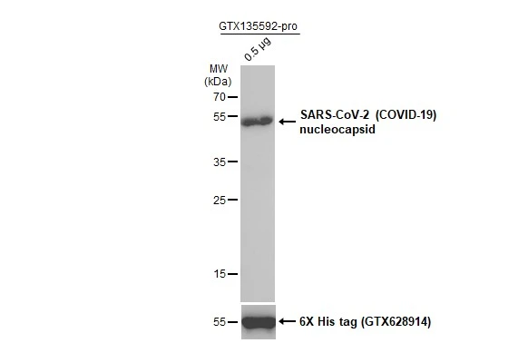
SARS-CoV-2 (COVID-19) nucleocapsid protein (GTX135592-pro, 0.5 μg) was separated by 12% SDS-PAGE, and the membrane was blotted with SARS-CoV / SARS-CoV-2 (COVID-19) nucleocapsid antibody [6H3] (GTX632269) diluted at 1:5000. The HRP-conjugated anti-mouset IgG antibody (GTX213111-01) was used to detect the primary antibody.
![Sandwich ELISA detection of non-transfected and SARS-CoV-2 nucleocapsid (full length) transfected 293T whole cell extracts using SARS-CoV / SARS-CoV-2 (COVID-19) nucleocapsid antibody [6H3] (GTX632269) as capture antibody at concentration of 5 弮g/mL and SARS-CoV-2 (COVID-19) nucleocapsid antibody (GTX135357) as detection antibody at concentration of 1 弮g/mL. Rabbit IgG antibody (HRP) (GTX213110-01) was diluted at 1:10000 and used to detect the primary antibody.](https://www.genetex.com/upload/website/prouct_img/normal/GTX632269/GTX632269_43936_20200605_ELISA_PAIR_B_w_23061202_385.webp "Sandwich ELISA detection of non-transfected and SARS-CoV-2 nucleocapsid (full length) transfected 293T whole cell extracts using SARS-CoV / SARS-CoV-2 (COVID-19) nucleocapsid antibody [6H3] (GTX632269) as capture antibody at concentration of 5 弮g/mL and SARS-CoV-2 (COVID-19) nucleocapsid antibody (GTX135357) as detection antibody at concentration of 1 弮g/mL. Rabbit IgG antibody (HRP) (GTX213110-01) was diluted at 1:10000 and used to detect the primary antibody.")
 nucleocapsid protein (GTX135357-pro) using GTX135361 as capture antibody at concentration of 5 μg/mL and GTX632269 as detection antibody at concentration of 1 μg/mL. Mouse IgG antibody (HRP) (GTX213111-01) was diluted at 1:10000 and used to detect the primary antibody.")
![Immunoprecipitation of SARS-CoV-2 NP transfected 293T whole cell extracts using 2 μg of SARS-CoV / SARS-CoV-2 (COVID-19) nucleocapsid antibody [6H3] (GTX632269). Western blot analysis was performed using SARS-CoV / SARS-CoV-2 (COVID-19) nucleocapsid antibody [6H3] (GTX632269). EasyBlot HRP-conjugated anti mouse IgG antibody (GTX221667-01) was used to detect the primary antibody.](https://www.genetex.com/upload/website/prouct_img/normal/GTX632269/GTX632269_42079_20200417_IP_B_w_23061202_484.webp "Immunoprecipitation of SARS-CoV-2 NP transfected 293T whole cell extracts using 2 μg of SARS-CoV / SARS-CoV-2 (COVID-19) nucleocapsid antibody [6H3] (GTX632269). Western blot analysis was performed using SARS-CoV / SARS-CoV-2 (COVID-19) nucleocapsid antibody [6H3] (GTX632269). EasyBlot HRP-conjugated anti mouse IgG antibody (GTX221667-01) was used to detect the primary antibody.")
![SARS-CoV / SARS-CoV-2 (COVID-19) nucleocapsid antibody [6H3] detects SARS-CoV-2 (COVID-19) nucleocapsid protein by immunohistochemical analysis. Sample: Mock (GTX435670) and SARS-CoV-2 (COVID-19) Nucleocapsid transfected 293T cell FFPE Cell Pellet Block (GTX435641). Red: SARS-CoV-2 (COVID-19) nucleocapsid stained by SARS-CoV / SARS-CoV-2 (COVID-19) nucleocapsid antibody [6H3] (GTX632269) diluted at 1:1000. Blue: Fluoroshield with DAPI (GTX30920). Antigen Retrieval: Citrate buffer, pH 6.0, 15 min](https://www.genetex.com/upload/website/prouct_img/normal/GTX632269/GTX632269_43936_20200515_IHC-P-FL_B_w_23061202_426.webp "SARS-CoV / SARS-CoV-2 (COVID-19) nucleocapsid antibody [6H3] detects SARS-CoV-2 (COVID-19) nucleocapsid protein by immunohistochemical analysis. Sample: Mock (GTX435670) and SARS-CoV-2 (COVID-19) Nucleocapsid transfected 293T cell FFPE Cell Pellet Block (GTX435641). Red: SARS-CoV-2 (COVID-19) nucleocapsid stained by SARS-CoV / SARS-CoV-2 (COVID-19) nucleocapsid antibody [6H3] (GTX632269) diluted at 1:1000. Blue: Fluoroshield with DAPI (GTX30920). Antigen Retrieval: Citrate buffer, pH 6.0, 15 min")
![Non-transfected (–) and transfected (+) 293T whole cell extracts (30 μg) were separated by 10% SDS-PAGE, and the membrane was blotted with SARS-CoV / SARS-CoV-2 (COVID-19) nucleocapsid antibody [6H3] (GTX632269) diluted at 1:5000. The HRP-conjugated anti-mouset IgG antibody (GTX213111-01) was used to detect the primary antibody, and the signal was developed with Trident ECL plus-Enhanced.](https://www.genetex.com/upload/website/prouct_img/normal/GTX632269/GTX632269_43936_20200526_WB_B_multiplevirus_w_23061202_516.webp "Non-transfected (–) and transfected (+) 293T whole cell extracts (30 μg) were separated by 10% SDS-PAGE, and the membrane was blotted with SARS-CoV / SARS-CoV-2 (COVID-19) nucleocapsid antibody [6H3] (GTX632269) diluted at 1:5000. The HRP-conjugated anti-mouset IgG antibody (GTX213111-01) was used to detect the primary antibody, and the signal was developed with Trident ECL plus-Enhanced.")
 nucleocapsid protein (GTX135357-pro) using GTX135357 as capture antibody at concentration of 5 μg/mL and GTX632269 as detection antibody at concentration of 1 μg/mL. Mouse IgG antibody (HRP) (GTX213111-01) was diluted at 1:10000 and used to detect the primary antibody.")
![SARS-CoV / SARS-CoV-2 (COVID-19) nucleocapsid antibody [6H3] detects SARS-CoV / SARS-CoV-2 (COVID-19) nucleocapsid protein by immunohistochemical analysis. Sample: Paraffin-embedded SARS-CoV-2 (COVID-19) Nucleocapsid FFPE Cell Pellet Block. Red: SARS-CoV / SARS-CoV-2 (COVID-19) nucleocapsid stained by SARS-CoV / SARS-CoV-2 (COVID-19) nucleocapsid antibody [6H3] (GTX632269) diluted at 1:1000. Green: SARS-CoV-2 (COVID-19) nucleocapsid stained by SARS-CoV-2 (COVID-19) nucleocapsid antibody (GTX135357) diluted at 1:1000. Blue: Fluoroshield with DAPI (GTX30920). Antigen Retrieval: Citrate buffer, pH 6.0, 15 min](https://www.genetex.com/upload/website/prouct_img/normal/GTX632269/GTX632269_43936_20200430_IHC-P-FL_Virus_2_w_23061202_770.webp "SARS-CoV / SARS-CoV-2 (COVID-19) nucleocapsid antibody [6H3] detects SARS-CoV / SARS-CoV-2 (COVID-19) nucleocapsid protein by immunohistochemical analysis. Sample: Paraffin-embedded SARS-CoV-2 (COVID-19) Nucleocapsid FFPE Cell Pellet Block. Red: SARS-CoV / SARS-CoV-2 (COVID-19) nucleocapsid stained by SARS-CoV / SARS-CoV-2 (COVID-19) nucleocapsid antibody [6H3] (GTX632269) diluted at 1:1000. Green: SARS-CoV-2 (COVID-19) nucleocapsid stained by SARS-CoV-2 (COVID-19) nucleocapsid antibody (GTX135357) diluted at 1:1000. Blue: Fluoroshield with DAPI (GTX30920). Antigen Retrieval: Citrate buffer, pH 6.0, 15 min")
![Non-infected (–) and infected (+, 48h pl MOI 0.01) Caco2 whole cell extracts were separated by SDS-PAGE, and the membrane was blotted with SARS-CoV / SARS-CoV-2 (COVID-19) nucleocapsid antibody [6H3] (GTX632269) diluted at 1:1000.](https://www.genetex.com/upload/website/prouct_img/normal/GTX632269/GTX632269_42079_20200305_WB_SARScoronavirus_w_23061202_857.webp "Non-infected (–) and infected (+, 48h pl MOI 0.01) Caco2 whole cell extracts were separated by SDS-PAGE, and the membrane was blotted with SARS-CoV / SARS-CoV-2 (COVID-19) nucleocapsid antibody [6H3] (GTX632269) diluted at 1:1000.")
![WB analysis of SARS-CoV infected Vero E6 cells harvested at different time-points post infection (T = 0, 8, 16 and 24 h) using GTX632269 SARS-CoV / SARS-CoV-2 (COVID-19) nucleocapsid antibody [6H3].](https://www.genetex.com/upload/website/prouct_img/normal/GTX632269/GTX632269_20200221_WB_w_23061202_994.webp "WB analysis of SARS-CoV infected Vero E6 cells harvested at different time-points post infection (T = 0, 8, 16 and 24 h) using GTX632269 SARS-CoV / SARS-CoV-2 (COVID-19) nucleocapsid antibody [6H3].")
SARS-CoV-2 (COVID-19) nucleocapsid protein (GTX135592-pro, 0.5 μg) was separated by 12% SDS-PAGE, and the membrane was blotted with SARS-CoV / SARS-CoV-2 (COVID-19) nucleocapsid antibody [6H3] (GTX632269) diluted at 1:5000. The HRP-conjugated anti-mouset IgG antibody (GTX213111-01) was used to detect the primary antibody.
SARS-CoV / SARS-CoV-2 (COVID-19) Nucleocapsid antibody [6H3]
GTX632269
ApplicationsImmunoFluorescence, ImmunoPrecipitation, Western Blot, ELISA, ImmunoCytoChemistry, ImmunoHistoChemistry, ImmunoHistoChemistry Frozen, ImmunoHistoChemistry Paraffin
Product group Antibodies
ReactivityVirus
Overview
- SupplierGeneTex
- Product NameSARS-CoV / SARS-CoV-2 (COVID-19) Nucleocapsid antibody [6H3]
- Delivery Days Customer9
- Application Supplier NoteWB: 1:1000-1:10000. *Optimal dilutions/concentrations should be determined by the researcher.Not tested in other applications.
- ApplicationsImmunoFluorescence, ImmunoPrecipitation, Western Blot, ELISA, ImmunoCytoChemistry, ImmunoHistoChemistry, ImmunoHistoChemistry Frozen, ImmunoHistoChemistry Paraffin
- CertificationResearch Use Only
- ClonalityMonoclonal
- Clone ID6H3
- Concentration1.5 mg/ml
- ConjugateUnconjugated
- HostMouse
- IsotypeIgG1
- ReactivityVirus
- Storage Instruction-20°C or -80°C,2°C to 8°C
- UNSPSC41116161