

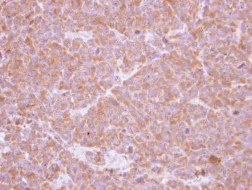
Immunohistochemical analysis of paraffin-embedded H1299 xenograft , using SBDS(GTX109168) antibody at 1:500 dilution.
Antigen Retrieval: Trilogy? (EDTA based, pH 8.0) buffer, 15min
 antibody at 1:500 dilution.")
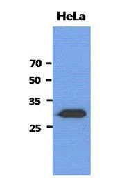
 A: H1299 B: HeLa C: Hep G2 (GTX27900) 12% SDS PAGE GTX109168 diluted at 1:1000")
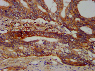
![SBDS antibody [N1C3] detects SBDS protein at cytosol on mouse duodenum by immunohistochemical analysis. Sample: Paraffin-embedded mouse duodenum. SBDS antibody [N1C3] (GTX109168) dilution: 1:500.
Antigen Retrieval: Trilogy? (EDTA based, pH 8.0) buffer, 15min](https://www.genetex.com/upload/website/prouct_img/normal/GTX109168/GTX109168_40023_IHC_M_w_23060120_382.webp "SBDS antibody [N1C3] detects SBDS protein at cytosol on mouse duodenum by immunohistochemical analysis. Sample: Paraffin-embedded mouse duodenum. SBDS antibody [N1C3] (GTX109168) dilution: 1:500.
Antigen Retrieval: Trilogy? (EDTA based, pH 8.0) buffer, 15min")
 A: mouse liver 12% SDS PAGE GTX109168 diluted at 1:1000")
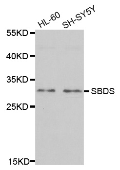
![Various whole cell extracts (30 μg) were separated by 12% SDS-PAGE, and the membrane was blotted with SBDS antibody [N1C3] (GTX109168) diluted at 1:1000. The HRP-conjugated anti-rabbit IgG antibody (GTX213110-01) was used to detect the primary antibody.](https://www.genetex.com/upload/website/prouct_img/normal/GTX109168/GTX109168_44447_20210924_WB_23102401_669.webp "Various whole cell extracts (30 μg) were separated by 12% SDS-PAGE, and the membrane was blotted with SBDS antibody [N1C3] (GTX109168) diluted at 1:1000. The HRP-conjugated anti-rabbit IgG antibody (GTX213110-01) was used to detect the primary antibody.")
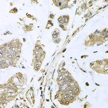
Immunohistochemical analysis of paraffin-embedded H1299 xenograft , using SBDS(GTX109168) antibody at 1:500 dilution.
Antigen Retrieval: Trilogy? (EDTA based, pH 8.0) buffer, 15min
SBDS antibody [N1C3]
GTX109168
ApplicationsImmunoFluorescence, ImmunoPrecipitation, Western Blot, ImmunoCytoChemistry, ImmunoHistoChemistry, ImmunoHistoChemistry Paraffin
Product group Antibodies
ReactivityHuman, Mouse
TargetSBDS
Overview
- SupplierGeneTex
- Product NameSBDS antibody [N1C3]
- Delivery Days Customer9
- Application Supplier NoteWB: 1:500-1:3000. ICC/IF: 1:100-1:1000. IHC-P: 1:100-1:1000. *Optimal dilutions/concentrations should be determined by the researcher.Not tested in other applications.
- ApplicationsImmunoFluorescence, ImmunoPrecipitation, Western Blot, ImmunoCytoChemistry, ImmunoHistoChemistry, ImmunoHistoChemistry Paraffin
- CertificationResearch Use Only
- ClonalityPolyclonal
- Concentration1.07 mg/ml
- ConjugateUnconjugated
- Gene ID51119
- Target nameSBDS
- Target descriptionSBDS ribosome maturation factor
- Target synonymsCGI-97, SDO1, SDS, SWDS, ribosome maturation protein SBDS, SBDS, ribosome assembly guanine nucleotide exchange factor
- HostRabbit
- IsotypeIgG
- Protein IDQ9Y3A5
- Protein NameRibosome maturation protein SBDS
- Scientific DescriptionThis gene encodes a member of a highly conserved protein family that exists from archaea to vertebrates and plants. The encoded protein may function in RNA metabolism. Mutations within this gene are associated with Shwachman-Bodian-Diamond syndrome. An alternative transcript has been described, but its biological nature has not been determined. This gene has a closely linked pseudogene that is distally located. [provided by RefSeq]
- ReactivityHuman, Mouse
- Storage Instruction-20°C or -80°C,2°C to 8°C
- UNSPSC41116161
Datasheet
Related products
Product group Antibodies
Anti-SBDS AntibodyA31237
ApplicationsWestern Blot, ImmunoHistoChemistry
ReactivityHuman, Mouse, Rat
- SizePrice

Product group Antibodies
Anti-SBDS Antibody Picoband(r)A02396-1-CARRIER-FREE
ApplicationsFlow Cytometry, ImmunoFluorescence, Western Blot, ELISA, ImmunoCytoChemistry
ReactivityHuman, Mouse, Rat
TargetSBDS
- SizePrice
Product group Antibodies
Anti-SBDS Antibody144-05876
ApplicationsImmunoFluorescence, Western Blot, ImmunoHistoChemistry
ReactivityHuman, Mouse, Rat
TargetSBDS
- SizePrice
Product group Antibodies
SBDS Monoclonal AntibodyBSM-60373M
ApplicationsImmunoFluorescence, Western Blot, ImmunoCytoChemistry, ImmunoHistoChemistry, ImmunoHistoChemistry Frozen, ImmunoHistoChemistry Paraffin
ReactivityHuman, Mouse, Rat
TargetSBDS
- SizePrice
Product group Antibodies
SBDS AntibodyCSB-PA897481LA01HU
ApplicationsImmunoFluorescence, ELISA, ImmunoHistoChemistry
ReactivityHuman
TargetSBDS
- SizePrice
Product group Antibodies

SBDS AntibodyLS-C334358
ApplicationsImmunoFluorescence, Western Blot, ImmunoHistoChemistry
ReactivityHuman, Mouse, Rat
TargetSBDS
- SizePrice

Product group Antibodies
Anti-SBDS AntibodyHPA028891
ApplicationsWestern Blot, ImmunoCytoChemistry, ImmunoHistoChemistry
ReactivityHuman, Mouse, Rat
TargetSBDS
- SizePrice
Product group Antibodies
SBDS antibodyGTX64702
ApplicationsImmunoFluorescence, Western Blot, ImmunoCytoChemistry, ImmunoHistoChemistry, ImmunoHistoChemistry Paraffin
ReactivityHuman, Mouse, Rat
TargetSBDS
- SizePrice
Product group Antibodies
SBDS antibody [AT1E8]GTX53781
ApplicationsWestern Blot, ELISA
ReactivityHuman
TargetSBDS
- SizePrice