

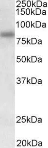
WB analysis of HEK293 cell lysate using GTX57169 SDCCAG8 antibody. Dilution : 0.3μg/ml Loading : 35μg protein in RIPA buffer

WB analysis of HEK293 cell lysate using GTX57169 SDCCAG8 antibody. Dilution : 0.3μg/ml Loading : 35μg protein in RIPA buffer
SDCCAG8 antibody
GTX57169
ApplicationsWestern Blot, ELISA, ImmunoHistoChemistry, ImmunoHistoChemistry Paraffin
Product group Antibodies
ReactivityHuman
TargetSDCCAG8
Overview
- SupplierGeneTex
- Product NameSDCCAG8 antibody
- Delivery Days Customer9
- Application Supplier NoteWB: 0.3-1microg/ml. IHC-P: 5microg/ml. ELISA: 1:128000. *Optimal dilutions/concentrations should be determined by the researcher.Not tested in other applications.
- ApplicationsWestern Blot, ELISA, ImmunoHistoChemistry, ImmunoHistoChemistry Paraffin
- CertificationResearch Use Only
- ClonalityPolyclonal
- Concentration0.50 mg/ml
- ConjugateUnconjugated
- Gene ID10806
- Target nameSDCCAG8
- Target descriptionSHH signaling and ciliogenesis regulator SDCCAG8
- Target synonymsBBS16, CCCAP, CCCAP SLSN7, HSPC085, NPHP10, NY-CO-8, SLSN7, hCCCAP, serologically defined colon cancer antigen 8, Bardet-Biedl syndrome 16, antigen NY-CO-8, centrosomal colon cancer autoantigen protein, nephrocystin 10
- HostGoat
- IsotypeIgG
- Scientific DescriptionThis gene encodes a centrosome associated protein. This protein may be involved in organizing the centrosome during interphase and mitosis. Mutations in this gene are associated with retinal-renal ciliopathy. [provided by RefSeq, Oct 2010]
- ReactivityHuman
- Storage Instruction-20°C or -80°C,2°C to 8°C
- UNSPSC41116161
Datasheet
Related products
Product group Antibodies
Anti-SDCCAG8 AntibodyA286070
ApplicationsELISA, ImmunoHistoChemistry
ReactivityHuman
- SizePrice
Product group Antibodies
Anti-SDCCAG8 (N-term) Antibody102-22757
ApplicationsWestern Blot
TargetSDCCAG8
- SizePrice
Product group Antibodies
SDCCAG8 Polyclonal AntibodyBS-7011R
ApplicationsImmunoFluorescence, Western Blot, ELISA, ImmunoCytoChemistry, ImmunoHistoChemistry, ImmunoHistoChemistry Frozen, ImmunoHistoChemistry Paraffin
ReactivityBovine, Equine, Human, Mouse, Porcine, Rat, Sheep
TargetSDCCAG8
- SizePrice
Product group Antibodies
SDCCAG8 AntibodyCSB-PA006722
ApplicationsELISA, ImmunoHistoChemistry
ReactivityHuman, Mouse
TargetSDCCAG8
- SizePrice
Product group Antibodies

Goat anti-SDCCAG8EB11471
ApplicationsWestern Blot, ELISA, ImmunoHistoChemistry
ReactivityCanine, Human, Mouse, Porcine, Rat
TargetSDCCAG8
- SizePrice
Product group Antibodies

ApplicationsELISA, ImmunoHistoChemistry
ReactivityHuman, Mouse
TargetSDCCAG8
- SizePrice

Product group Antibodies
Anti-SDCCAG8 AntibodyHPA025737
ApplicationsImmunoCytoChemistry
ReactivityHuman
TargetSDCCAG8
- SizePrice
![SDCCAG8 antibody detects SDCCAG8 protein at centrosome by immunofluorescent analysis. Sample: A549 cells were fixed in ice-cold MeOH for 5 min. Green: SDCCAG8 protein stained by SDCCAG8 antibody (GTX119588) diluted at 1:200. Red: alpha Tubulin, a cytoskeleton marker, stained by alpha Tubulin antibody [GT114] diluted at 1:1000. Blue: Hoechst 33342 staining. Scale bar = 10 μm.](https://www.genetex.com/upload/website/prouct_img/normal/GTX119588/GTX119588_41185_IFA_w_23060519_448.webp)
Product group Antibodies
SDCCAG8 antibodyGTX119588
ApplicationsImmunoFluorescence, ImmunoCytoChemistry
ReactivityHuman
TargetSDCCAG8
- SizePrice
Product group Antibodies
SDCCAG8 antibody, InternalGTX45798
ApplicationsWestern Blot
ReactivityHuman
TargetSDCCAG8
- SizePrice
Product group Antibodies
SDCCAG8 antibody, N-termGTX45799
ApplicationsWestern Blot
ReactivityHuman
TargetSDCCAG8
- SizePrice