

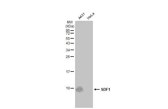
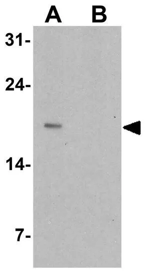
Various extracts (30 μg) were separated by 15% SDS-PAGE, and the membrane was blotted with SDF1 antibody [N1C3] (GTX116092) diluted at 1:1000. The HRP-conjugated anti-rabbit IgG antibody (GTX213110-01) was used to detect the primary antibody.
![SDF1 antibody [N1C3] detects SDF1 protein by immunofluorescent analysis. Sample: DIV9 rat cortical neuron and Glia cell cells were fixed in 4% paraformaldehyde at RT for 15 min. Green: SDF1 stained by SDF1 antibody [N1C3] (GTX116092) diluted at 1:50. Red: Tau, an axon marker, stained by Tau antibody [GT287] (GTX634809) diluted at 1:500.](https://www.genetex.com/upload/website/prouct_img/normal/GTX116092/GTX116092_44419_20211112_ICC_IF_R_w_23060519_321.webp "SDF1 antibody [N1C3] detects SDF1 protein by immunofluorescent analysis. Sample: DIV9 rat cortical neuron and Glia cell cells were fixed in 4% paraformaldehyde at RT for 15 min. Green: SDF1 stained by SDF1 antibody [N1C3] (GTX116092) diluted at 1:50. Red: Tau, an axon marker, stained by Tau antibody [GT287] (GTX634809) diluted at 1:500.")
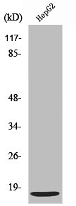
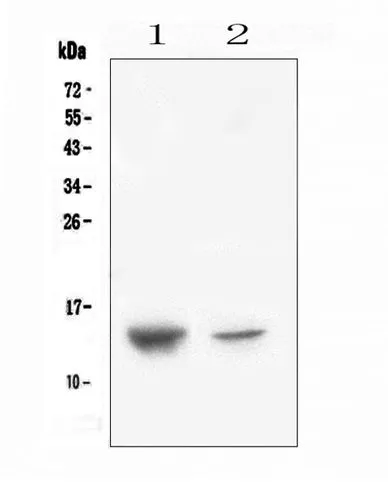
![Whole cell extract (30 μg) was separated by 15% SDS-PAGE, and the membrane was blotted with SDF1 antibody [N1C3] (GTX116092) diluted at 1:1000. The HRP-conjugated anti-rabbit IgG antibody (GTX213110-01) was used to detect the primary antibody.](https://www.genetex.com/upload/website/prouct_img/normal/GTX116092/GTX116092_41724_20200918_WB_2_w_23060519_708.webp "Whole cell extract (30 μg) was separated by 15% SDS-PAGE, and the membrane was blotted with SDF1 antibody [N1C3] (GTX116092) diluted at 1:1000. The HRP-conjugated anti-rabbit IgG antibody (GTX213110-01) was used to detect the primary antibody.")

Various extracts (30 μg) were separated by 15% SDS-PAGE, and the membrane was blotted with SDF1 antibody [N1C3] (GTX116092) diluted at 1:1000. The HRP-conjugated anti-rabbit IgG antibody (GTX213110-01) was used to detect the primary antibody.
SDF1 antibody [N1C3]
GTX116092
ApplicationsImmunoFluorescence, Western Blot, ImmunoCytoChemistry
Product group Antibodies
ReactivityHuman, Mouse, Rat
TargetCXCL12
Overview
- SupplierGeneTex
- Product NameSDF1 antibody [N1C3]
- Delivery Days Customer9
- Application Supplier NoteWB: 1:500-1:3000. *Optimal dilutions/concentrations should be determined by the researcher.Not tested in other applications.
- ApplicationsImmunoFluorescence, Western Blot, ImmunoCytoChemistry
- CertificationResearch Use Only
- ClonalityPolyclonal
- Concentration0.16 mg/ml
- ConjugateUnconjugated
- Gene ID6387
- Target nameCXCL12
- Target descriptionC-X-C motif chemokine ligand 12
- Target synonymsIRH, PBSF, SCYB12, SDF1, TLSF, TPAR1, stromal cell-derived factor 1, chemokine (C-X-C motif) ligand 12, intercrine reduced in hepatomas, pre-B cell growth-stimulating factor
- HostRabbit
- IsotypeIgG
- Protein IDP48061
- Protein NameStromal cell-derived factor 1
- Scientific DescriptionFor background information on chemokines, see CXCL1 (MIM 155730). Stromal cell-derived factors 1-alpha and 1-beta are small cytokines that belong to the intercrine family, members of which activate leukocytes and are often induced by proinflammatory stimuli such as lipopolysaccharide, TNF (see MIM 191160), or IL1 (see MIM 147760). The intercrines are characterized by the presence of 4 conserved cysteines which form 2 disulfide bonds. They can be classified into 2 subfamilies. In the CC subfamily, which includes beta chemokine, the cysteine residues are adjacent to each other. In the CXC subfamily, which includes alpha chemokine, they are separated by an intervening amino acid. The SDF1 proteins belong to the latter group.[supplied by OMIM]
- ReactivityHuman, Mouse, Rat
- Storage Instruction-20°C or -80°C,2°C to 8°C
- UNSPSC41116161
Datasheet
Related products
Product group Antibodies
Anti-CXCL12 Antibody144-62714
ApplicationsImmunoFluorescence, Western Blot, ImmunoHistoChemistry
ReactivityHuman, Mouse, Rat
TargetCXCL12
- SizePrice

Product group Antibodies
Anti-CXCL12 Antibody Picoband(r)A00053-1-CARRIER-FREE
ApplicationsWestern Blot, ImmunoHistoChemistry
ReactivityMouse, Rat
TargetCXCL12
- SizePrice
Product group Antibodies
CXCL12 Polyclonal AntibodyBS-4938R
ApplicationsImmunoFluorescence, Western Blot, ELISA, ImmunoCytoChemistry, ImmunoHistoChemistry, ImmunoHistoChemistry Frozen, ImmunoHistoChemistry Paraffin
ReactivityCanine, Chicken, Guinea Pig, Human, Mouse, Porcine, Rabbit, Rat
TargetCXCL12
- SizePrice
Product group Antibodies
CXCL12 AntibodyCSB-PA004056
ApplicationsWestern Blot, ELISA, ImmunoHistoChemistry
ReactivityHuman, Mouse, Rat
TargetCXCL12
- SizePrice
Product group Antibodies
ApplicationsImmunoPrecipitation, Western Blot, ImmunoCytoChemistry, ImmunoHistoChemistry
ReactivityMouse, Rat
TargetCXCL12
- SizePrice
Product group Antibodies
SDF1 antibodyGTX31900
ApplicationsImmunoFluorescence, Western Blot, ELISA, ImmunoCytoChemistry
ReactivityHuman, Mouse
TargetCXCL12
- SizePrice
Product group Antibodies
SDF1 antibodyGTX02876
ApplicationsWestern Blot, ELISA, ImmunoHistoChemistry, ImmunoHistoChemistry Paraffin
ReactivityHuman, Mouse, Rat
TargetCXCL12
- SizePrice
Product group Antibodies
SDF1 antibodyGTX44804
ApplicationsImmunoPrecipitation, Western Blot, ImmunoHistoChemistry
ReactivityMouse
TargetCXCL12
- SizePrice
Product group Antibodies
SDF1 antibody [9N26]GTX52664
ApplicationsFlow Cytometry, ImmunoHistoChemistry, ImmunoHistoChemistry Paraffin
ReactivityHuman
TargetCXCL12
- SizePrice