

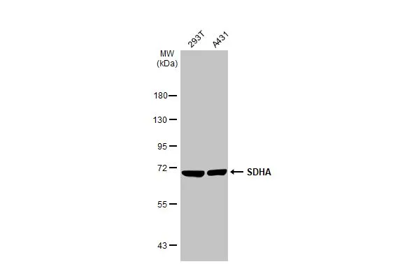
Various whole cell extracts (30 μg) were separated by 7.5% SDS-PAGE, and the membrane was blotted with SDHA antibody [GT20710] (GTX632636) diluted at 1:3000. The HRP-conjugated anti-mouse IgG antibody (GTX213111-01) was used to detect the primary antibody.
![SDHA antibody [GT20710] detects SDHA protein by immunohistochemical analysis. Sample: Paraffin-embedded mouse tissues. SDHA stained by SDHA antibody [GT20710] (GTX632636) diluted at 1:200. Antigen Retrieval: Citrate buffer, pH 6.0, 15 min](https://www.genetex.com/upload/website/prouct_img/normal/GTX632636/GTX632636_44748_20230512_IHC-P_multiple_M_23060622_630.webp "SDHA antibody [GT20710] detects SDHA protein by immunohistochemical analysis. Sample: Paraffin-embedded mouse tissues. SDHA stained by SDHA antibody [GT20710] (GTX632636) diluted at 1:200. Antigen Retrieval: Citrate buffer, pH 6.0, 15 min")
![SDHA antibody [GT20710] detects SDHA protein at mitochondria by immunohistochemical analysis. Sample: Paraffin-embedded mouse kidney. SDHA stained by SDHA antibody [GT20710] (GTX632636) diluted at 1:200. Antigen Retrieval: Citrate buffer, pH 6.0, 15 min](https://www.genetex.com/upload/website/prouct_img/normal/GTX632636/GTX632636_42702_20181207_IHC-P_M_1_w_23061202_766.webp "SDHA antibody [GT20710] detects SDHA protein at mitochondria by immunohistochemical analysis. Sample: Paraffin-embedded mouse kidney. SDHA stained by SDHA antibody [GT20710] (GTX632636) diluted at 1:200. Antigen Retrieval: Citrate buffer, pH 6.0, 15 min")
![SDHA antibody [GT20710] detects SDHA protein at mitochondria by immunohistochemical analysis. Sample: Paraffin-embedded mouse liver. SDHA stained by SDHA antibody [GT20710] (GTX632636) diluted at 1:200. Antigen Retrieval: Citrate buffer, pH 6.0, 15 min](https://www.genetex.com/upload/website/prouct_img/normal/GTX632636/GTX632636_42702_20181207_IHC-P_M_w_23061202_804.webp "SDHA antibody [GT20710] detects SDHA protein at mitochondria by immunohistochemical analysis. Sample: Paraffin-embedded mouse liver. SDHA stained by SDHA antibody [GT20710] (GTX632636) diluted at 1:200. Antigen Retrieval: Citrate buffer, pH 6.0, 15 min")
![SDHA antibody [GT20710] detects SDHA protein at mitochondria by immunofluorescent analysis. Sample: HeLa cells were fixed in 4% paraformaldehyde at RT for 15 min. Green: SDHA protein stained by SDHA antibody [GT20710] (GTX632636) diluted at 1:500. Red: Phalloidin, a cytoskeleton marker, diluted at 1:100. Blue: Hoechst 33342 staining.](https://www.genetex.com/upload/website/prouct_img/normal/GTX632636/GTX632636_42702_20170315_IFA_w_23061202_858.webp "SDHA antibody [GT20710] detects SDHA protein at mitochondria by immunofluorescent analysis. Sample: HeLa cells were fixed in 4% paraformaldehyde at RT for 15 min. Green: SDHA protein stained by SDHA antibody [GT20710] (GTX632636) diluted at 1:500. Red: Phalloidin, a cytoskeleton marker, diluted at 1:100. Blue: Hoechst 33342 staining.")
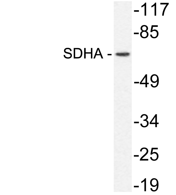
![Various whole cell extracts (30 μg) were separated by 7.5% SDS-PAGE, and the membrane was blotted with SDHA antibody [GT20710] (GTX632636) diluted at 1:3000.](https://www.genetex.com/upload/website/prouct_img/normal/GTX632636/GTX632636_42702_20170119_WB_M_w_23061202_734.webp "Various whole cell extracts (30 μg) were separated by 7.5% SDS-PAGE, and the membrane was blotted with SDHA antibody [GT20710] (GTX632636) diluted at 1:3000.")
![Non-transfected (–) and transfected (+) 293T whole cell extracts (30 μg) were separated by 7.5% SDS-PAGE, and the membrane was blotted with SDHA antibody [GT20710] (GTX632636) diluted at 1:1000.](https://www.genetex.com/upload/website/prouct_img/normal/GTX632636/GTX632636_42289_20160825_WB_shRNA_watermark_w_23061202_574.webp "Non-transfected (–) and transfected (+) 293T whole cell extracts (30 μg) were separated by 7.5% SDS-PAGE, and the membrane was blotted with SDHA antibody [GT20710] (GTX632636) diluted at 1:1000.")
![Various whole cell extracts (30 μg) were separated by 7.5% SDS-PAGE, and the membrane was blotted with SDHA antibody [GT20710] (GTX632636) diluted at 1:3000.](https://www.genetex.com/upload/website/prouct_img/normal/GTX632636/GTX632636_42702_20170119_WB_R_w_23061202_875.webp "Various whole cell extracts (30 μg) were separated by 7.5% SDS-PAGE, and the membrane was blotted with SDHA antibody [GT20710] (GTX632636) diluted at 1:3000.")
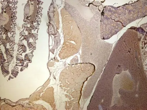
![SDHA antibody [GT20710] detects SDHA protein at mitochondria by immunohistochemical analysis. Sample: Paraffin-embedded rat colon. SDHA stained by SDHA antibody [GT20710] (GTX632636) diluted at 1:200. Antigen Retrieval: Citrate buffer, pH 6.0, 15 min](https://www.genetex.com/upload/website/prouct_img/normal/GTX632636/GTX632636_42702_20181207_IHC-P_R_w_23061202_655.webp "SDHA antibody [GT20710] detects SDHA protein at mitochondria by immunohistochemical analysis. Sample: Paraffin-embedded rat colon. SDHA stained by SDHA antibody [GT20710] (GTX632636) diluted at 1:200. Antigen Retrieval: Citrate buffer, pH 6.0, 15 min")
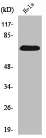
![HepG2 and mitochondria extracts (30 μg) were separated by SDS-PAGE, and the membrane was blotted with SDHA antibody [GT20710] (GTX632636) diluted at 1:1000. The HRP-conjugated anti-mouse IgG antibody (GTX213111-01) was used to detect the primary antibody.](https://www.genetex.com/upload/website/prouct_img/normal/GTX632636/GTX632636_42702_20191129_WB_Fraction_w_23061202_265.webp "HepG2 and mitochondria extracts (30 μg) were separated by SDS-PAGE, and the membrane was blotted with SDHA antibody [GT20710] (GTX632636) diluted at 1:1000. The HRP-conjugated anti-mouse IgG antibody (GTX213111-01) was used to detect the primary antibody.")
Various whole cell extracts (30 μg) were separated by 7.5% SDS-PAGE, and the membrane was blotted with SDHA antibody [GT20710] (GTX632636) diluted at 1:3000. The HRP-conjugated anti-mouse IgG antibody (GTX213111-01) was used to detect the primary antibody.
SDHA antibody [GT20710]
GTX632636
ApplicationsImmunoFluorescence, Western Blot, ImmunoCytoChemistry, ImmunoHistoChemistry, ImmunoHistoChemistry Paraffin
Product group Antibodies
ReactivityHuman, Mouse, Rat
TargetSDHA
Overview
- SupplierGeneTex
- Product NameSDHA antibody [GT20710]
- Delivery Days Customer9
- Application Supplier NoteWB: 1:500-1:3000. ICC/IF: 1:100-1:1000. IHC-P: 1:100-1:1000. *Optimal dilutions/concentrations should be determined by the researcher.Not tested in other applications.
- ApplicationsImmunoFluorescence, Western Blot, ImmunoCytoChemistry, ImmunoHistoChemistry, ImmunoHistoChemistry Paraffin
- CertificationResearch Use Only
- ClonalityMonoclonal
- Clone IDGT20710
- Concentration1 mg/ml
- ConjugateUnconjugated
- Gene ID6389
- Target nameSDHA
- Target descriptionsuccinate dehydrogenase complex flavoprotein subunit A
- Target synonymsCMD1GG, FP, MC2DN1, NDAXOA, PGL5, PPGL5, SDH1, SDH2, SDHF, succinate dehydrogenase [ubiquinone] flavoprotein subunit, mitochondrial, flavoprotein subunit of complex II, malate dehydrogenase [quinone] flavoprotein subunit, succinate dehydrogenase complex, subunit A, flavoprotein (Fp)
- HostMouse
- IsotypeIgG1
- Protein IDP31040
- Protein NameSuccinate dehydrogenase [ubiquinone] flavoprotein subunit, mitochondrial
- Scientific DescriptionThis gene encodes a major catalytic subunit of succinate-ubiquinone oxidoreductase, a complex of the mitochondrial respiratory chain. The complex is composed of four nuclear-encoded subunits and is localized in the mitochondrial inner membrane. Mutations in this gene have been associated with a form of mitochondrial respiratory chain deficiency known as Leigh Syndrome. A pseudogene has been identified on chromosome 3q29. [provided by RefSeq]
- ReactivityHuman, Mouse, Rat
- Storage Instruction-20°C or -80°C,2°C to 8°C
- UNSPSC41116161
Datasheet
Related products
Product group Antibodies
Anti-SDHA AntibodyA97917
ApplicationsWestern Blot, ELISA
ReactivityHuman, Mouse, Rat
- SizePrice
Product group Antibodies
Anti-SDHA Antibody144-02594
ApplicationsImmunoFluorescence, Western Blot, ImmunoHistoChemistry
ReactivityHuman, Mouse, Rat
TargetSDHA
- SizePrice

Product group Antibodies
Anti-SDHA Antibody Picoband(r)A01753-CARRIER-FREE
ApplicationsFlow Cytometry, ImmunoFluorescence, Western Blot, ImmunoCytoChemistry
ReactivityHuman, Mouse, Rat
TargetSDHA
- SizePrice
Product group Antibodies
SDHA Recombinant Antibody, AbBy Fluor-350 ConjugatedBSM-61435R-BF350
ApplicationsFlow Cytometry, ImmunoFluorescence, Western Blot
ReactivityHuman, Mouse, Rat
TargetSDHA
- SizePrice
Product group Antibodies
SDHA AntibodyCSB-PA004057
ApplicationsWestern Blot, ELISA
ReactivityHuman, Mouse, Rat
TargetSDHA
- SizePrice
Product group Antibodies
SDHA Polyclonal AntibodyCAC13909
ApplicationsImmunoFluorescence, Western Blot, ELISA, ImmunoHistoChemistry
TargetSDHA
- SizePrice
Product group Antibodies
SDHA antibodyGTX101689
ApplicationsImmunoFluorescence, Western Blot, ImmunoCytoChemistry, ImmunoHistoChemistry, ImmunoHistoChemistry Paraffin
ReactivityHuman, Mouse, Rat, Zebra Fish
TargetSDHA
- SizePrice

Product group Antibodies
Anti-SDHA AntibodyHPA041981
ApplicationsWestern Blot, ImmunoCytoChemistry, ImmunoHistoChemistry
ReactivityHuman
TargetSDHA
- SizePrice
![IHC-P analysis of human gastric antrum tissue using GTX640916 SDHA antibody [HMV336] HistoMAX?. A granular cytoplasmic SDHA staining occurs in all cells but it is strongest in stomach glands, especially in parietal cells.](https://www.genetex.com/upload/website/prouct_img/normal/GTX640916/GTX640916_20241015_IHC-P_2_24101420_507.webp)
Product group Antibodies
SDHA antibody [HMV336] HistoMAX(tm)GTX640916
ApplicationsImmunoHistoChemistry, ImmunoHistoChemistry Paraffin
ReactivityHuman
TargetSDHA
- SizePrice
![Various whole cell extracts (30 μg) were separated by 7.5% SDS-PAGE, and the membrane was blotted with SDHA antibody [HL1067] (GTX636098) diluted at 1:500. The HRP-conjugated anti-rabbit IgG antibody (GTX213110-01) was used to detect the primary antibody.](https://www.genetex.com/upload/website/prouct_img/normal/GTX636098/GTX636098_44438_20221028_WB_22110201_379.webp)
Product group Antibodies
SDHA antibody [HL1067]GTX636098
ApplicationsImmunoFluorescence, Western Blot, ImmunoCytoChemistry
ReactivityDrosophila, Human, Mouse, Rat, Zebra Fish
TargetSDHA
- SizePrice