

Search results: AP2M1
Product group Antibodies
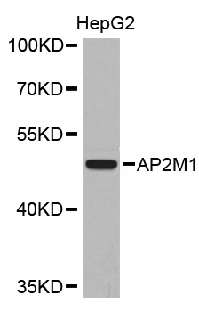
Anti-AP2M1 AntibodyA30558
ApplicationsWestern Blot, ImmunoHistoChemistry
ReactivityHuman, Mouse, Rat
- SizePrice
Product group Antibodies
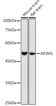
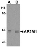
Ap2M1 Polyclonal AntibodyCAC07420
ApplicationsWestern Blot, ELISA, ImmunoHistoChemistry
TargetAP2M1
- SizePrice
Product group Antibodies
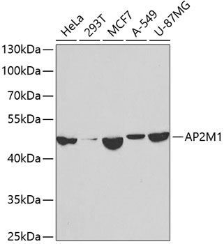
Anti-AP2M1 Antibody144-02492
ApplicationsWestern Blot, ImmunoHistoChemistry
ReactivityHuman, Mouse, Rat
TargetAP2M1
- SizePrice
Product group Antibodies
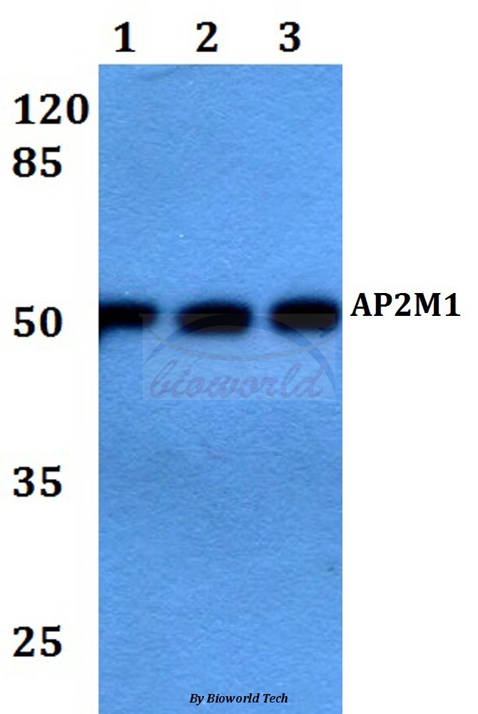
Anti-AP2M1 Antibody107-10265
ApplicationsWestern Blot, ImmunoHistoChemistry, ImmunoHistoChemistry Paraffin
ReactivityHuman
TargetAP2M1
- SizePrice
Product group Antibodies
Anti-AP2M1 AntibodyA92117
ApplicationsWestern Blot
ReactivityHuman, Mouse, Rat
- SizePrice

Product group Antibodies
Anti-AP2M1 AntibodyA37590
ApplicationsWestern Blot, ImmunoHistoChemistry
ReactivityHuman, Mouse, Rat
- SizePrice
Product group Antibodies
AP2M1 Polyclonal AntibodyCAC13020
ApplicationsWestern Blot, ELISA, ImmunoHistoChemistry
ReactivityMouse, Rat
TargetAP2M1
- SizePrice
Product group Antibodies
Anti-AP2M1 AntibodyA28295
ApplicationsWestern Blot
ReactivityHuman, Mouse, Rat
- SizePrice
Product group Antibodies
Anti-AP2M1 AntibodyA283441
ApplicationsWestern Blot
ReactivityHuman, Mouse, Rat
- SizePrice
Product group Antibodies
Anti-AP2M1 AntibodyA47995
ApplicationsWestern Blot, ELISA, ImmunoHistoChemistry
ReactivityHuman, Mouse, Rat
- SizePrice
Product group Antibodies
AP2M1 Recombinant AntibodyBSM-61305R
ApplicationsWestern Blot
TargetAP2M1
- SizePrice
Didn't find what you were looking for?
Search through our product groups to find the right product
Back to overview