Bio-Connect

Search results: CD115

541 to 552 of 615 products
Sort by
Western blot analysis of lysate from U-937 cell line, using CSF1R Antibody (Cat. #TA324948). TA324948 was diluted at 1:1000. A goat anti-rabbit IgG H&L (HRP) at 1:5000 dilution was used as the secondary antibody. Lysate at 35ug.
Product group Antibodies
OriGene
ApplicationsFlow Cytometry, ImmunoFluorescence, Western Blot
ReactivityHuman
TargetCSF1R
  • SizePrice
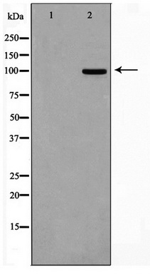
Western blot analysis on 293 cell lysate using M-CSF Receptor Antibody
Product group Antibodies
OriGene
ApplicationsWestern Blot
ReactivityHuman, Mouse, Rat
TargetCSF1R
  • SizePrice
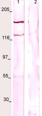
WB: The whole cell lysate derived from m-CSF stimulated LNCaP was immunoblotted by Rabbit anti-CSF-1R (pY723) at 1:500. A major immune-reactive band is observed around ~150kDa (lane 1). This band is blocked by pre-incubation of immunizing peptide (lane 2)
Product group Antibodies
OriGene
ApplicationsWestern Blot
ReactivityHuman, Mouse, Rat
TargetCSF1R
  • SizePrice
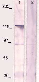
WB: The whole cell lysate derived from LNCaP was immunoblotted by Rabbit anti-CSF-1R (Paired Y723) at 1:500. An immunoreactive band is observed around ~130kDa (lane 1). This band is blocked by pre-incubation of immunizing peptide (lane 2)
Product group Antibodies
OriGene
ApplicationsWestern Blot
ReactivityHuman, Mouse, Rat
TargetCSF1R
  • SizePrice
WB: The whole cell lysate derived from LNCaP was immunoblotted by Rabbit anti-CSF-1R at 1:500. A major immunoreactive band is observed around ~130kDa (lane 1). This band is blocked by pre-incubation of immunizing peptide (lane 2)
Product group Antibodies
OriGene
ApplicationsWestern Blot
ReactivityHuman, Mouse, Rat
TargetCSF1R
  • SizePrice
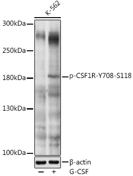
Western blot analysis of extracts of K-562 cells, using Phospho-CSF1R-Y708 antibody (TA375132) at 1:1000 dilution.K-562 cells were treated by G-CSF (25 ng/ml) at 37°C for 30 minutes after serum-starvation overnight. - Secondary antibody: HRP Goat Anti-Rabbit IgG (H+L) at 1:10000 dilution. - Lysates/proteins: 25ug per lane. - Blocking buffer: 3% nonfat dry milk in TBST. - Detection: ECL Basic Kit . - Exposure time: 1s.
Product group Antibodies
OriGene
ApplicationsWestern Blot
ReactivityHuman
TargetCSF1R
  • SizePrice
Western blot analysis of anti-CSF1R Pab (Cat. #TA300025) in human placenta. CSF1R (arrow) was detected using purified Pab. Secondary HRP-anti-rabbit was used for signal visualization with chemiluminescence.
Product group Antibodies
OriGene
ApplicationsFlow Cytometry, Western Blot, ImmunoHistoChemistry
ReactivityHuman
TargetCSF1R
  • SizePrice
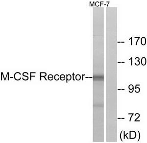
Western blot analysis of extracts from MCF-7 cells, using M-CSF Receptor (Ab-723) antibody.The lane on the right is treated with the synthesized peptide.
Product group Antibodies
OriGene
ApplicationsWestern Blot, ImmunoHistoChemistry
ReactivityHuman, Mouse
TargetCSF1R
  • SizePrice
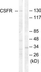
Western blot analysis of extracts from HT-29 cells, using CSFR (Ab-809) antibody.The lane on the right is treated with the synthesized peptide.
Product group Antibodies
OriGene
ApplicationsImmunoFluorescence, Western Blot, ImmunoHistoChemistry
ReactivityHuman, Mouse
TargetCSF1R
  • SizePrice
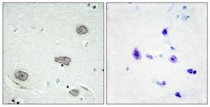
Immunohistochemistry analysis of paraffin-embedded human brain tissue using CSFR (Phospho-Tyr809) antibody.The picture on the right is treated with the synthesized peptide.
Product group Antibodies
OriGene
ApplicationsImmunoHistoChemistry
ReactivityHuman
TargetCSF1R
  • SizePrice
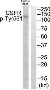
Western blot analysis of extracts from HepG2 cells treated with PMA, using CSFR (Phospho-Tyr561) antibody.The lane on the right is treated with the synthesized peptide.
Product group Antibodies
OriGene
ApplicationsWestern Blot, ImmunoHistoChemistry
ReactivityHuman, Mouse
TargetCSF1R
  • SizePrice
Product group Antibodies
OriGene
ApplicationsWestern Blot
ReactivityHuman
TargetCSF1R
  • SizePrice