

Search results: CDC25B
Product group Antibodies
ApplicationsWestern Blot
ReactivityHuman, Mouse, Rat
- SizePrice
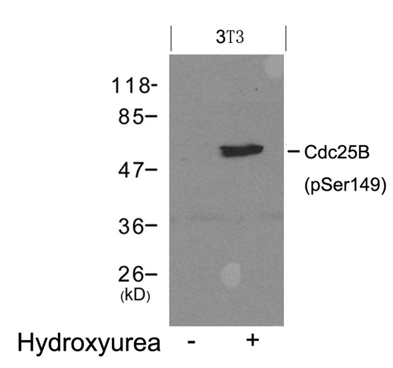
Product group Antibodies
ApplicationsWestern Blot
ReactivityMouse
- SizePrice
Product group Antibodies
ApplicationsWestern Blot
ReactivityHuman, Mouse
- SizePrice

Product group Antibodies
ApplicationsImmunoPrecipitation, Western Blot
ReactivityHuman
TargetCDC25B
- SizePrice

Product group Antibodies
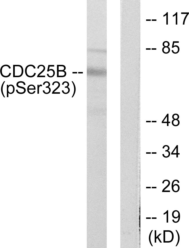
Anti-Phospho-Cdc25B (S323) AntibodyA01899S323
ApplicationsImmunoFluorescence, Western Blot, ELISA, ImmunoHistoChemistry
ReactivityHuman, Mouse, Rat
TargetCDC25B
- SizePrice
Product group Antibodies
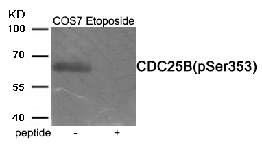
Anti-Phospho-Cdc25B (S353) AntibodyA01899S353
ApplicationsImmunoFluorescence, Western Blot, ELISA, ImmunoHistoChemistry
ReactivityHuman, Monkey
TargetCDC25B
- SizePrice
Product group Assays
Assay Sample TypeResearchers plate their cell line of choice
ReactivityHuman, Mouse, Rat
- SizePrice
Product group Antibodies
Anti-Cdc25B Rabbit Monoclonal AntibodyM01899-30UL
ApplicationsImmunoPrecipitation, Western Blot
ReactivityHuman
TargetCDC25B
- SizePrice
Product group Chemicals
CDC25B-IN-1 [2374831-10-2]HY-126246
CAS Number2374831-10-2
Estimated Purity98.12
Molecular Weight321.37
- SizePrice
![CDC25B-IN-1 [2374831-10-2]](https://www.targetmol.com/group3/M00/37/E3/CgoaEGayUy2Ea9oXAAAAAOFxg5Q334.png)
Product group Chemicals
CAS Number2374831-10-2
Estimated Purity97.86%
Molecular Weight321.37
- SizePrice
Didn't find what you were looking for?
Search through our product groups to find the right product
Back to overview