

Search results: EMP2
Product group Proteins / Signaling Molecules
TMED9, Human (HEK293, His)HY-P76678
Protein IDQ9BVK6
- SizePrice
Product group Proteins / Signaling Molecules
TMED9, Human (HEK293, Fc)HY-P76679
Protein IDQ9BVK6
- SizePrice
Product group Proteins / Signaling Molecules
Protein IDQ13445
- SizePrice
Product group Proteins / Signaling Molecules
Protein IDQ13445
- SizePrice
Product group Proteins / Signaling Molecules
TMED1, Human (HEK293, Fc)HY-P72445
Protein IDQ13445
- SizePrice
Product group Proteins / Signaling Molecules
TMED1, Human (HEK293, His)HY-P77240
Protein IDQ13445
- SizePrice
Product group Proteins / Signaling Molecules
TMED4, Human (HEK293, His)HY-P77242
Protein IDQ7Z7H5
- SizePrice
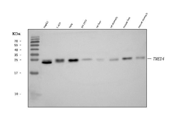
Product group Antibodies
Anti-TMED4 Antibody Picoband(r)A12647-1-BIOTIN
ApplicationsWestern Blot
ReactivityHuman, Mouse, Rat
TargetTMED4
- SizePrice
Product group Antibodies
Anti-TMED4 Antibody Picoband(r)A12647-1-CARRIER-FREE
ApplicationsWestern Blot
ReactivityHuman, Mouse, Rat
TargetTMED4
- SizePrice
Product group Antibodies
Anti-TMED4 Antibody Picoband(r)A12647-1-CY3
ApplicationsWestern Blot
ReactivityHuman, Mouse, Rat
TargetTMED4
- SizePrice
Product group Antibodies
Anti-TMED4 Antibody Picoband(r)A12647-1-DYLIGHT488
ApplicationsWestern Blot
ReactivityHuman, Mouse, Rat
TargetTMED4
- SizePrice
Product group Antibodies
Anti-TMED4 Antibody Picoband(r)A12647-1-DYLIGHT550
ApplicationsWestern Blot
ReactivityHuman, Mouse, Rat
TargetTMED4
- SizePrice
Didn't find what you were looking for?
Search through our product groups to find the right product
Back to overview