

Search results: FGF9
Product group Antibodies
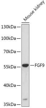
FGF9 antibodyGTX55621
ApplicationsWestern Blot
ReactivityHuman, Mouse
- SizePrice
Product group Antibodies
FGF9 antibodyGTX74042
ApplicationsWestern Blot, ELISA, Neutralisation/Blocking
ReactivityMouse
- SizePrice
Product group Antibodies
FGF9 AntibodyLS-C756543
ApplicationsWestern Blot
ReactivityHuman, Mouse, Rat
- SizePrice
Product group Antibodies
FGF9 AntibodyLS-C742922
ApplicationsWestern Blot, ELISA
ReactivityHuman, Mouse, Rat
- SizePrice
Product group Antibodies
FGF9 AntibodyLS-C809987
ApplicationsWestern Blot, ELISA, ImmunoHistoChemistry
ReactivityHuman, Mouse, Rat
- SizePrice

Product group Antibodies
FGF9 antibody [4D25]GTX52745
ApplicationsWestern Blot, ImmunoHistoChemistry
ReactivityHuman
- SizePrice


Product group Antibodies
Anti-FGF9HPA067007
ApplicationsImmunoHistoChemistry
ReactivityHuman
TargetFGF9
- SizePrice
Didn't find what you were looking for?
Search through our product groups to find the right product
Back to overview