

Search results: HAUS8
Product group Antibodies
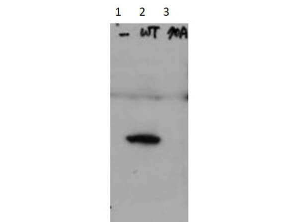
HAUS8 Rabbit Polyclonal AntibodyTA397437S
ApplicationsWestern Blot, ELISA
ReactivityHuman
TargetHAUS8
- SizePrice
Product group Antibodies
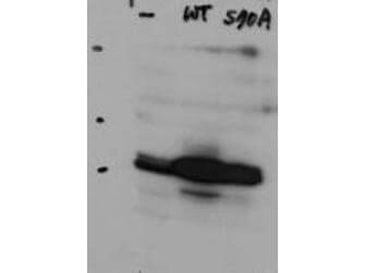
HAUS8 Rabbit Polyclonal AntibodyTA397438S
ApplicationsWestern Blot, ELISA
ReactivityHuman
TargetHAUS8
- SizePrice

Product group Antibodies
HAUS8 Rabbit Polyclonal AntibodyTA342771
ApplicationsWestern Blot
ReactivityHuman
TargetHAUS8
- SizePrice
Product group Antibodies
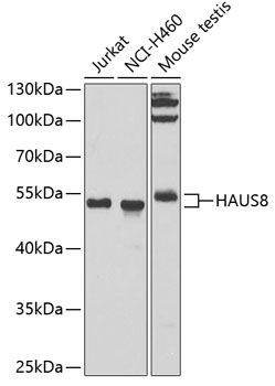
HAUS8 Rabbit Polyclonal AntibodyTA376930
ApplicationsImmunoFluorescence, Western Blot, ImmunoCytoChemistry
ReactivityHuman, Mouse
TargetHAUS8
- SizePrice
Product group Lysates / Extracts

- SizePrice
Product group Proteins / Signaling Molecules
HAUS8 Peptide - N-terminal regionORB2010421
ApplicationsWestern Blot
Protein IDQ9BT25
- SizePrice
Product group Proteins / Signaling Molecules
HAUS8 Human Over-expression LysateORB1372364
Protein IDQ9BT25
- SizePrice
Product group Proteins / Signaling Molecules
HAUS8 Human Over-expression LysateORB1352028
Protein IDQ9BT25
- SizePrice
Didn't find what you were looking for?
Search through our product groups to find the right product
Back to overview