

Search results: JUP
Product group Antibodies
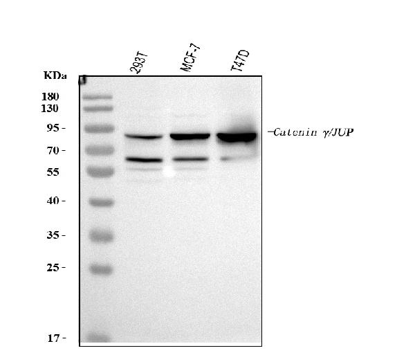
Anti-gamma Catenin/JUP Antibody Picoband(r)PA1117-1-10UG
ApplicationsFlow Cytometry, ImmunoFluorescence, Western Blot, ImmunoCytoChemistry
ReactivityHamster, Human
TargetJUP
- SizePrice
Product group Antibodies
Anti-gamma Catenin/JUP Antibody Picoband(r)PA1117-1-APC
ApplicationsFlow Cytometry, ImmunoFluorescence, Western Blot, ImmunoCytoChemistry
ReactivityHamster, Human
TargetJUP
- SizePrice
Product group Antibodies
Anti-gamma Catenin/JUP Antibody Picoband(r)PA1117-1-BIOTIN
ApplicationsFlow Cytometry, ImmunoFluorescence, Western Blot, ImmunoCytoChemistry
ReactivityHamster, Human
TargetJUP
- SizePrice
Product group Antibodies
Anti-gamma Catenin/JUP Antibody Picoband(r)PA1117-1-CARRIER-FREE
ApplicationsFlow Cytometry, ImmunoFluorescence, Western Blot, ImmunoCytoChemistry
ReactivityHamster, Human
TargetJUP
- SizePrice
Product group Antibodies
Anti-gamma Catenin/JUP Antibody Picoband(r)PA1117-1-CY3
ApplicationsFlow Cytometry, ImmunoFluorescence, Western Blot, ImmunoCytoChemistry
ReactivityHamster, Human
TargetJUP
- SizePrice
Product group Antibodies
Anti-gamma Catenin/JUP Antibody Picoband(r)PA1117-1-DYLIGHT488
ApplicationsFlow Cytometry, ImmunoFluorescence, Western Blot, ImmunoCytoChemistry
ReactivityHamster, Human
TargetJUP
- SizePrice
Product group Antibodies
Anti-gamma Catenin/JUP Antibody Picoband(r)PA1117-1-DYLIGHT550
ApplicationsFlow Cytometry, ImmunoFluorescence, Western Blot, ImmunoCytoChemistry
ReactivityHamster, Human
TargetJUP
- SizePrice
Product group Antibodies
Anti-gamma Catenin/JUP Antibody Picoband(r)PA1117-1-DYLIGHT594
ApplicationsFlow Cytometry, ImmunoFluorescence, Western Blot, ImmunoCytoChemistry
ReactivityHamster, Human
TargetJUP
- SizePrice
Product group Antibodies
Anti-gamma Catenin/JUP Antibody Picoband(r)PA1117-1-FITC
ApplicationsFlow Cytometry, ImmunoFluorescence, Western Blot, ImmunoCytoChemistry
ReactivityHamster, Human
TargetJUP
- SizePrice
Product group Antibodies
Anti-gamma Catenin/JUP Antibody Picoband(r)PA1117-1-HRP
ApplicationsFlow Cytometry, ImmunoFluorescence, Western Blot, ImmunoCytoChemistry
ReactivityHamster, Human
TargetJUP
- SizePrice
Product group Antibodies
Anti-gamma Catenin/JUP Antibody Picoband(r)PA1117-1-IFLUOR647
ApplicationsFlow Cytometry, ImmunoFluorescence, Western Blot, ImmunoCytoChemistry
ReactivityHamster, Human
TargetJUP
- SizePrice
Didn't find what you were looking for?
Search through our product groups to find the right product
Back to overview