

Search results: LOX CRISPR
Product group Antibodies
Lox Antibody, Biotin conjugatedCSB-PA013038LD01RA
ApplicationsELISA
ReactivityRat
TargetLox
- SizePrice
Product group Proteins / Signaling Molecules
Eukaryotic Lysyl Oxidase (LOX)EPC580HU61
ApplicationsWestern Blot, Other Application
Protein IDP28300
- SizePrice
Product group Antibodies

Lysyl Oxidase (LOX) AntibodyABX110154
ApplicationsWestern Blot, ELISA, ImmunoHistoChemistry
- SizePrice
Product group Antibodies
Lysyl Oxidase (LOX) AntibodyABX110470
ApplicationsWestern Blot, ELISA
- SizePrice
Product group Antibodies
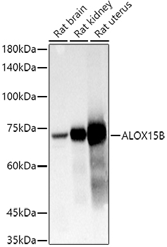
Anti-15-LOX-2 AntibodyA12029
ApplicationsImmunoFluorescence, Western Blot, ImmunoCytoChemistry
ReactivityHuman, Mouse, Rat
- SizePrice
Product group Proteins / Signaling Molecules
ApplicationsOther Application
- SizePrice
Product group Proteins / Signaling Molecules
Active Lysyl Oxidase (LOX)APC580HU01
ApplicationsFunctional Assay, Other Application
Protein IDP28300
- SizePrice
Product group Antibodies
Lysyl Oxidase (LOX) AntibodyABX214882
ApplicationsELISA, ImmunoHistoChemistry
- SizePrice
Product group Antibodies
Lysyl Oxidase (LOX) AntibodyABX241109
ApplicationsWestern Blot, ELISA, ImmunoHistoChemistry
- SizePrice
Product group Antibodies
Lysyl Oxidase (LOX) AntibodyABX241110
ApplicationsWestern Blot, ELISA, ImmunoHistoChemistry
- SizePrice
Product group Antibodies
lysyl Oxidase (LOX) AntibodyABX432943
ApplicationsWestern Blot, ELISA
- SizePrice
Didn't find what you were looking for?
Search through our product groups to find the right product
Back to overview