

Search results: LOX CRISPR
Product group Molecular Biology
- SizePrice
Product group Molecular Biology
- SizePrice
Product group Chemicals
5-LOX-IN-4 [125235-15-6]HY-U00308
CAS Number125235-15-6
Estimated Purity98.06
Molecular Weight424.56
- SizePrice
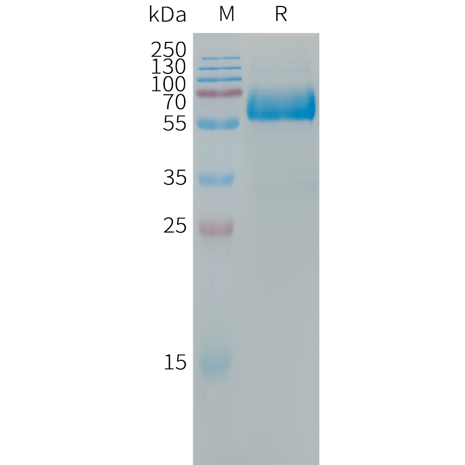
Product group Proteins / Signaling Molecules
ApplicationsOther Application
- SizePrice

Product group Antibodies
Anti-LOX 1/Olr1 Antibody Picoband(r)A00760-2-10UG
ApplicationsWestern Blot, ELISA
ReactivityMouse, Rat
TargetOlr1
- SizePrice
Product group Antibodies
Anti-LOX 1/Olr1 Antibody Picoband(r)A00760-2-APC
ApplicationsWestern Blot, ELISA
ReactivityMouse, Rat
TargetOlr1
- SizePrice
Product group Antibodies
Anti-LOX 1/Olr1 Antibody Picoband(r)A00760-2-BIOTIN
ApplicationsWestern Blot, ELISA
ReactivityMouse, Rat
TargetOlr1
- SizePrice
Product group Antibodies
Anti-LOX 1/Olr1 Antibody Picoband(r)A00760-2-CARRIER-FREE
ApplicationsWestern Blot, ELISA
ReactivityMouse, Rat
TargetOlr1
- SizePrice
Product group Antibodies
Anti-LOX 1/Olr1 Antibody Picoband(r)A00760-2-CY3
ApplicationsWestern Blot, ELISA
ReactivityMouse, Rat
TargetOlr1
- SizePrice
Product group Antibodies
Anti-LOX 1/Olr1 Antibody Picoband(r)A00760-2-DYLIGHT488
ApplicationsWestern Blot, ELISA
ReactivityMouse, Rat
TargetOlr1
- SizePrice
Didn't find what you were looking for?
Search through our product groups to find the right product
Back to overview