

Search results: NeuroD1
Product group Antibodies
NeuroD1 antibody, PAbORB33617
ApplicationsWestern Blot, ELISA
ReactivityHuman, Mouse, Rat
- SizePrice
Product group Antibodies
ApplicationsImmunoFluorescence, Western Blot, ELISA, ImmunoCytoChemistry, ImmunoHistoChemistry, ImmunoHistoChemistry Frozen, ImmunoHistoChemistry Paraffin
ReactivityHuman, Mouse, Rat
- SizePrice
Product group Antibodies
References
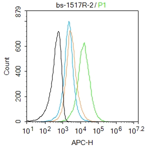
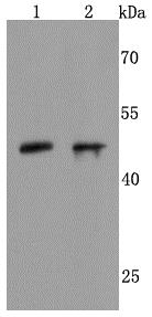
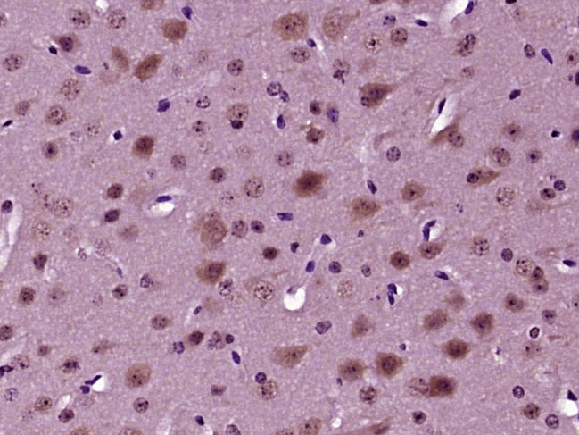
NeuroD1 Polyclonal AntibodyBS-1517R
ApplicationsFlow Cytometry, ImmunoFluorescence, Western Blot, ELISA, ImmunoCytoChemistry, ImmunoHistoChemistry, ImmunoHistoChemistry Frozen, ImmunoHistoChemistry Paraffin
ReactivityBovine, Canine, Human, Mouse, Porcine, Rat
- SizePrice
Product group Proteins / Signaling Molecules
Recombinant Human NeuroD1MBS7610427
Protein IDQ13562
- SizePrice

Product group Antibodies
NeuroD1 (1C9) Monoclonal AntibodyBSM-52948R
ApplicationsWestern Blot, ImmunoHistoChemistry, ImmunoHistoChemistry Paraffin
ReactivityHuman, Mouse, Rat
- SizePrice
Product group Antibodies
NeuroD1 (Ser274) Polyclonal AntibodyBS-19218R
ApplicationsImmunoFluorescence, Western Blot, ELISA, ImmunoCytoChemistry, ImmunoHistoChemistry, ImmunoHistoChemistry Frozen, ImmunoHistoChemistry Paraffin
ReactivityBovine, Human, Mouse, Porcine, Rat, Sheep
- SizePrice
Didn't find what you were looking for?
Search through our product groups to find the right product
Back to overview