

Search results: PRR5
Product group Antibodies
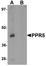
Anti-PRR5 AntibodyA49693
ApplicationsWestern Blot, ELISA
ReactivityHuman, Mouse, Rat
- SizePrice


Product group Antibodies
Anti-PRR5 AntibodyHPA054072
ApplicationsImmunoHistoChemistry
ReactivityHuman
TargetPRR5
- SizePrice

Product group Antibodies
Anti-PRR5 Antibody Picoband(r)A05975-1-10UG
ApplicationsFlow Cytometry, Western Blot, ELISA
ReactivityHuman
TargetPRR5
- SizePrice
Product group Antibodies
Anti-PRR5 Antibody Picoband(r)A05975-1-APC
ApplicationsFlow Cytometry, Western Blot, ELISA
ReactivityHuman
TargetPRR5
- SizePrice
Product group Antibodies
Anti-PRR5 Antibody Picoband(r)A05975-1-BIOTIN
ApplicationsFlow Cytometry, Western Blot, ELISA
ReactivityHuman
TargetPRR5
- SizePrice
Product group Antibodies
Anti-PRR5 Antibody Picoband(r)A05975-1-CARRIER-FREE
ApplicationsFlow Cytometry, Western Blot, ELISA
ReactivityHuman
TargetPRR5
- SizePrice
Product group Antibodies
Anti-PRR5 Antibody Picoband(r)A05975-1-CY3
ApplicationsFlow Cytometry, Western Blot, ELISA
ReactivityHuman
TargetPRR5
- SizePrice
Product group Antibodies
Anti-PRR5 Antibody Picoband(r)A05975-1-DYLIGHT488
ApplicationsFlow Cytometry, Western Blot, ELISA
ReactivityHuman
TargetPRR5
- SizePrice
Didn't find what you were looking for?
Search through our product groups to find the right product
Back to overview