

Search results: SIRT4

Product group Antibodies
Anti-SIRT4 AntibodyHPA029691
ApplicationsWestern Blot, ImmunoHistoChemistry
ReactivityHuman
TargetSIRT4
- SizePrice

Product group Antibodies
Anti-SIRT4 AntibodyHPA029692
ApplicationsWestern Blot, ImmunoHistoChemistry
ReactivityHuman
TargetSIRT4
- SizePrice
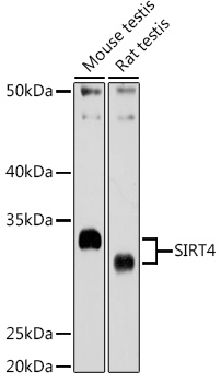
Product group Antibodies
Anti-SIRT4 AntibodyA12140
ApplicationsWestern Blot, ImmunoHistoChemistry
ReactivityHuman, Mouse, Rat
- SizePrice
Product group Antibodies
Anti-SIRT4 AntibodyER-14-1310
ApplicationsWestern Blot, ImmunoHistoChemistry, ImmunoHistoChemistry Paraffin
ReactivityBovine, Canine, Human, Mouse, Porcine, Rat
- SizePrice
Product group Proteins / Signaling Molecules

Human Sirtuin 4 (SIRT4) ProteinABX166511
ApplicationsWestern Blot, Other Application
- SizePrice
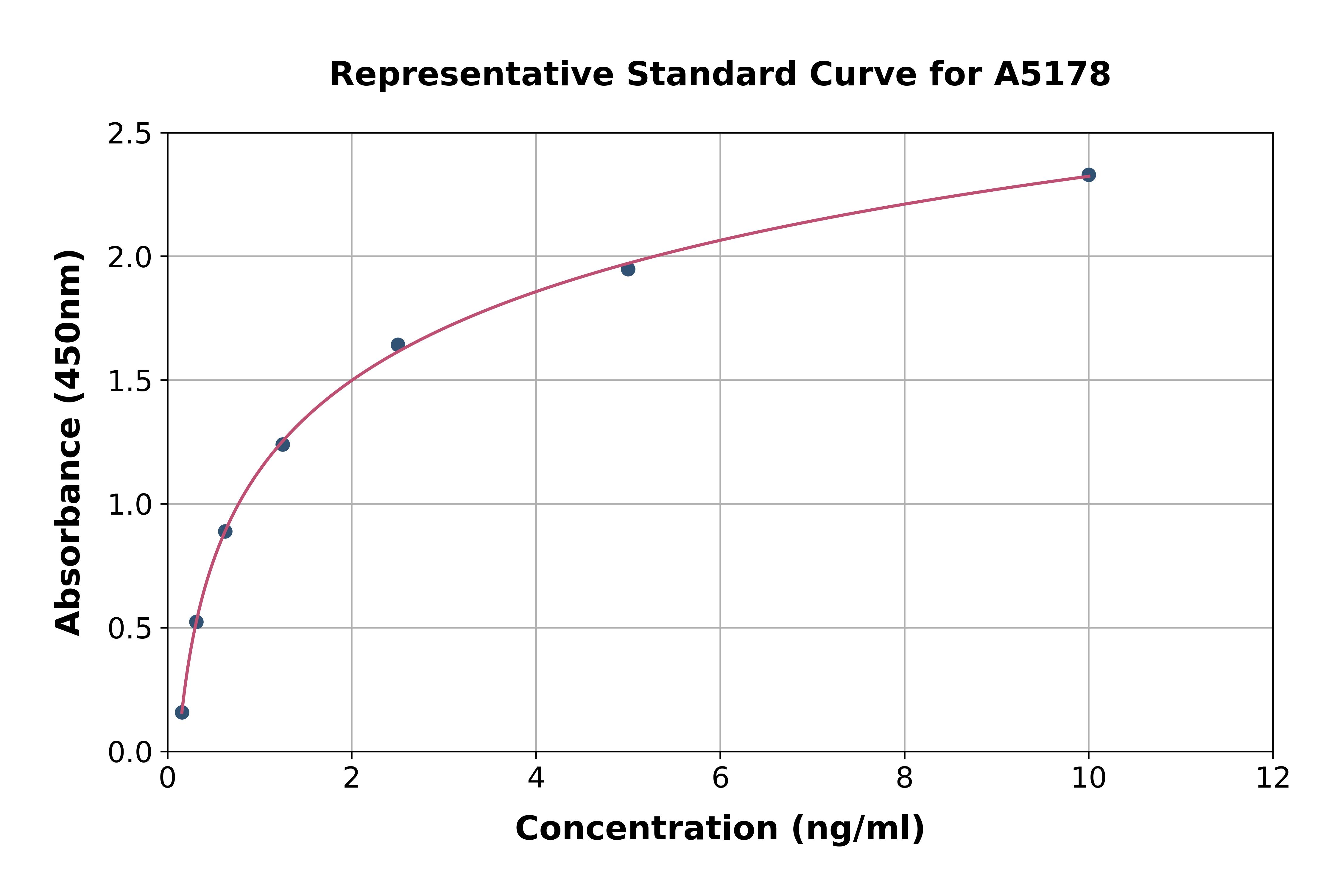
Product group Assays
Assay Sample TypeSerum, plasma, tissue homogenates, cell lysates, cell culture supernates and other biological fluids.
ReactivityHuman
- SizePrice
Product group Proteins / Signaling Molecules
Mouse Sirtuin 4 (SIRT4) ProteinABX069104
ApplicationsWestern Blot, Other Application
- SizePrice
Product group Proteins / Signaling Molecules
Rat Sirtuin 4 (SIRT4) ProteinABX069105
ApplicationsWestern Blot, Other Application
- SizePrice
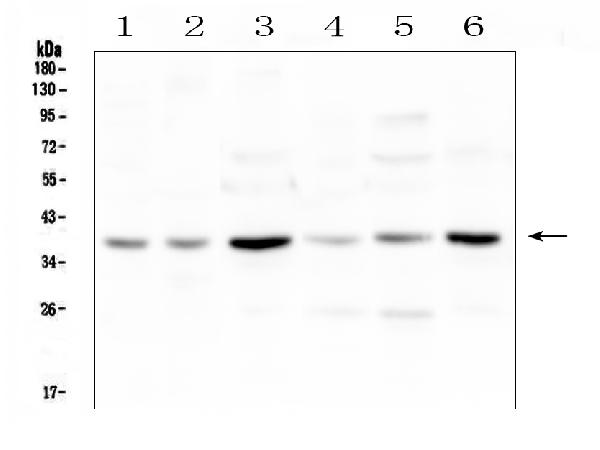
Product group Antibodies
Anti-SIRT4 Antibody Picoband(r)A03764-1-10UG
ApplicationsWestern Blot, ImmunoHistoChemistry
ReactivityHuman, Mouse, Rat
TargetSIRT4
- SizePrice
Didn't find what you were looking for?
Search through our product groups to find the right product
Back to overview