

Search results: Siglec-3

Product group Antibodies
ApplicationsImmunoPrecipitation, Western Blot, ImmunoHistoChemistry
ReactivityHuman, Mouse, Rat
TargetCSNK2B
- SizePrice
Product group Antibodies
ApplicationsImmunoPrecipitation, Western Blot, ImmunoHistoChemistry
ReactivityHuman, Mouse, Rat
TargetCSNK2B
- SizePrice

Product group Antibodies
Anti-CD52 Monoclonal AntibodyM03484-30UL
ApplicationsImmunoPrecipitation, Western Blot
ReactivityMouse
TargetCd52
- SizePrice
Product group Antibodies
ApplicationsImmunoPrecipitation, Western Blot
ReactivityMouse
TargetCd52
- SizePrice
Product group Antibodies
ApplicationsImmunoFluorescence, ImmunoPrecipitation, Western Blot, ImmunoCytoChemistry, ImmunoHistoChemistry
ReactivityHuman, Rat
TargetCRYAB
- SizePrice
Product group Antibodies
ApplicationsImmunoFluorescence, ImmunoPrecipitation, Western Blot, ImmunoCytoChemistry, ImmunoHistoChemistry
ReactivityHuman, Rat
TargetCRYAB
- SizePrice

Product group Antibodies
Anti-CCR8 Rabbit Monoclonal AntibodyM03518-30UL
ApplicationsImmunoFluorescence, Western Blot, ImmunoCytoChemistry
ReactivityHuman, Mouse, Rat
TargetCCR8
- SizePrice
Product group Antibodies
ApplicationsImmunoFluorescence, Western Blot, ImmunoCytoChemistry
ReactivityHuman, Mouse, Rat
TargetCCR8
- SizePrice
Product group Antibodies
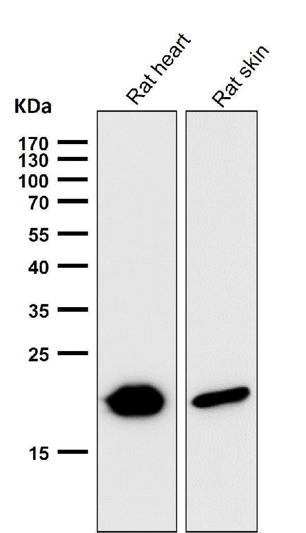
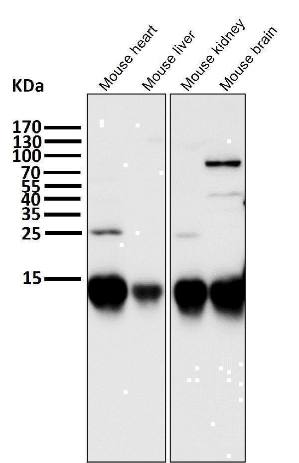
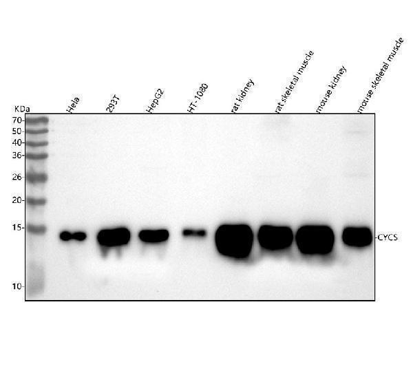
Anti-Cytochrome C CYCS Rabbit Monoclonal AntibodyM03529-1-30UL
ApplicationsWestern Blot, ImmunoHistoChemistry
ReactivityHuman, Mouse, Rat
TargetCYCS
- SizePrice
Product group Antibodies
ApplicationsWestern Blot, ImmunoHistoChemistry
ReactivityHuman, Mouse, Rat
TargetCYCS
- SizePrice
Product group Antibodies
Anti-Cytochrome C CYCS Rabbit Monoclonal AntibodyM03529-2-30UL
ApplicationsImmunoFluorescence, ImmunoPrecipitation, Western Blot, ImmunoCytoChemistry, ImmunoHistoChemistry
ReactivityHuman, Mouse, Rat
TargetCYCS
- SizePrice
Product group Antibodies
ApplicationsImmunoFluorescence, ImmunoPrecipitation, Western Blot, ImmunoCytoChemistry, ImmunoHistoChemistry
ReactivityHuman, Mouse, Rat
TargetCYCS
- SizePrice
Didn't find what you were looking for?
Search through our product groups to find the right product
Back to overview