

Search results: TCP1


Product group Antibodies
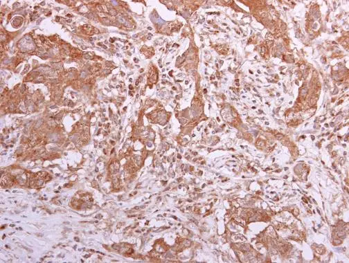
TCP1 alpha antibody, C-termGTX10180
ApplicationsWestern Blot, ImmunoHistoChemistry, ImmunoHistoChemistry Paraffin
ReactivityHuman
- SizePrice
Product group Antibodies
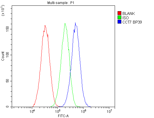
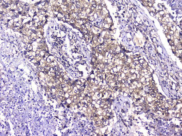
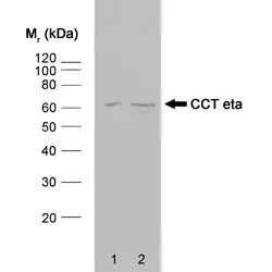
TCP1 eta antibody [PK/16/81a]GTX75919
ApplicationsWestern Blot, ImmunoHistoChemistry, ImmunoHistoChemistry Paraffin
ReactivityBovine, Human, Mouse, Rabbit
- SizePrice
Product group Antibodies
ApplicationsWestern Blot, ImmunoHistoChemistry, ImmunoHistoChemistry Paraffin
ReactivityBovine, Canine, Equine, Guinea Pig, Hamster, Human, Mammals, Monkey, Mouse, Porcine, Rabbit, Rat
- SizePrice
Didn't find what you were looking for?
Search through our product groups to find the right product
Back to overview