

Search results: USP2
Product group Antibodies
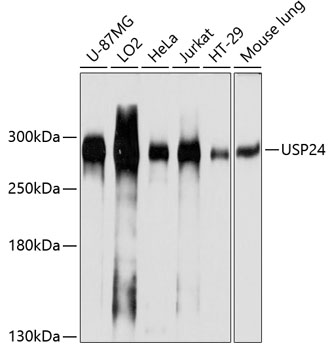
USP24 Recombinant AntibodyBSM-62458R
ApplicationsWestern Blot
TargetUSP24
- SizePrice
Product group Antibodies
Usp24 Recombinant AntibodyCAC12652
ApplicationsFlow Cytometry, ImmunoFluorescence, ELISA
TargetUSP24
- SizePrice
Product group Antibodies
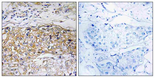
Anti-USP24 AntibodyA08241-2
ApplicationsImmunoFluorescence, Western Blot, ELISA, ImmunoCytoChemistry, ImmunoHistoChemistry
- SizePrice
Product group Antibodies
Anti-USP24 AntibodyA08241
ApplicationsImmunoFluorescence, ELISA, ImmunoCytoChemistry, ImmunoHistoChemistry
TargetUSP24
- SizePrice
Product group Antibodies
Anti-USP24 AntibodyA04288-1
ApplicationsImmunoFluorescence, Western Blot, ImmunoHistoChemistry
- SizePrice

Product group DNA / RNA / Vectors
Human, USP25 cDNA ORF Clone, untaggedHG20539-UT
CategoryDNA / RNA / Vectors
- SizePrice
Product group Antibodies
USP28 AntibodyCSB-PA025718GA01HU
ApplicationsWestern Blot, ELISA, ImmunoHistoChemistry
TargetUSP28
- SizePrice
Didn't find what you were looking for?
Search through our product groups to find the right product
Back to overview