

Search results: USP2

Product group Antibodies
Anti-USP26 Antibody Picoband(r)A07732-1-CY3
ApplicationsFlow Cytometry, Western Blot, ELISA, ImmunoHistoChemistry
TargetUSP26
- SizePrice
Product group Antibodies
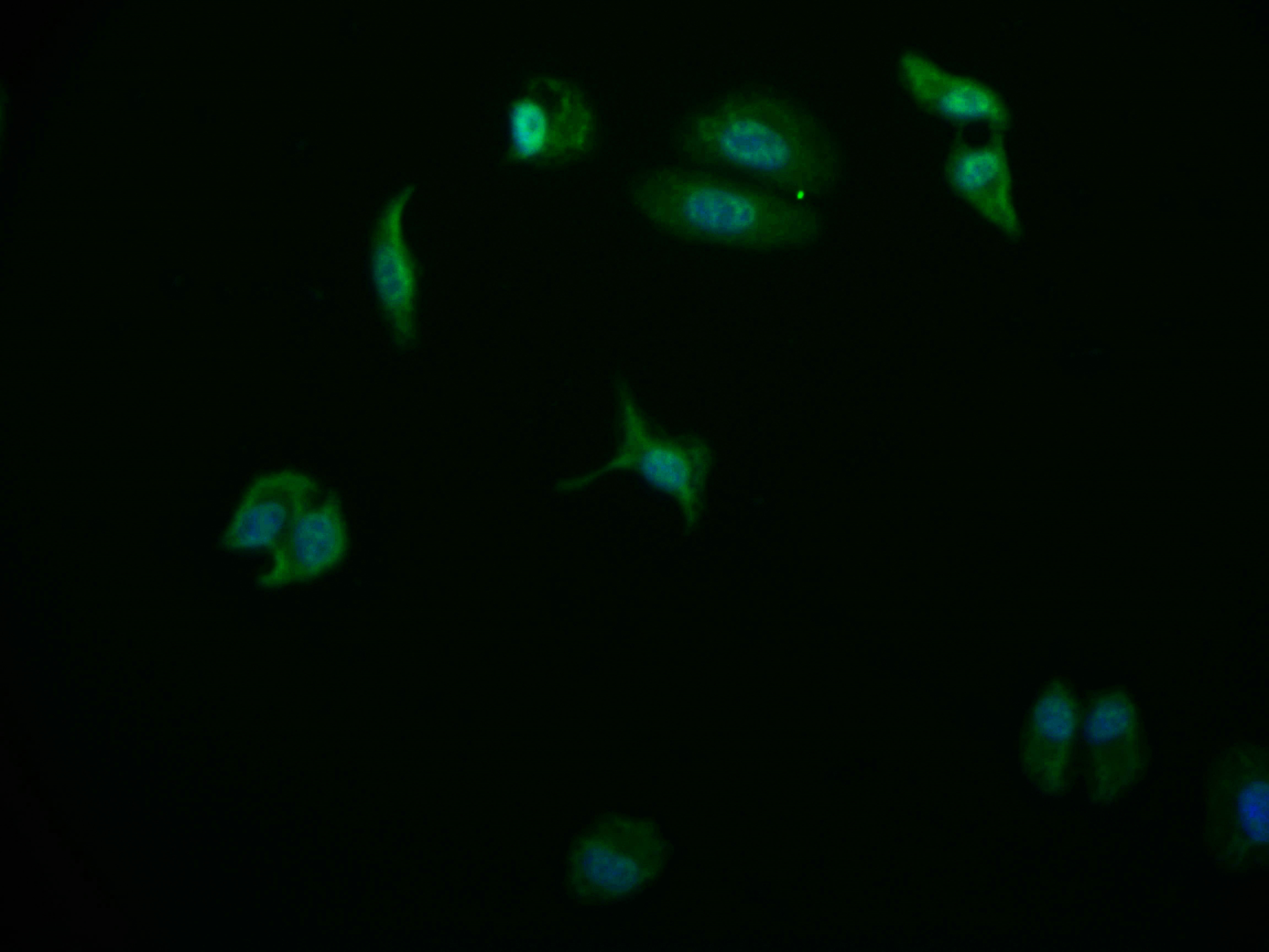
Anti-USP26 Antibody Picoband(r)A07732-1-DYLIGHT488
ApplicationsFlow Cytometry, Western Blot, ELISA, ImmunoHistoChemistry
TargetUSP26
- SizePrice
Product group Antibodies
Anti-USP26 Antibody Picoband(r)A07732-1-DYLIGHT550
ApplicationsFlow Cytometry, Western Blot, ELISA, ImmunoHistoChemistry
TargetUSP26
- SizePrice
Product group Antibodies
Anti-USP26 Antibody Picoband(r)A07732-1-DYLIGHT594
ApplicationsFlow Cytometry, Western Blot, ELISA, ImmunoHistoChemistry
TargetUSP26
- SizePrice
Product group Antibodies
Anti-USP26 Antibody Picoband(r)A07732-1-FITC
ApplicationsFlow Cytometry, Western Blot, ELISA, ImmunoHistoChemistry
TargetUSP26
- SizePrice
Product group Antibodies
Anti-USP26 Antibody Picoband(r)A07732-1-HRP
ApplicationsFlow Cytometry, Western Blot, ELISA, ImmunoHistoChemistry
TargetUSP26
- SizePrice
Product group Antibodies
Anti-USP26 Antibody Picoband(r)A07732-1-IFLUOR647
ApplicationsFlow Cytometry, Western Blot, ELISA, ImmunoHistoChemistry
TargetUSP26
- SizePrice
Product group Antibodies
Anti-USP26 Antibody Picoband(r)A07732-1-PE
ApplicationsFlow Cytometry, Western Blot, ELISA, ImmunoHistoChemistry
TargetUSP26
- SizePrice
Product group Antibodies
Anti-USP26 Antibody Picoband(r)A07732-1
ApplicationsFlow Cytometry, Western Blot, ELISA, ImmunoHistoChemistry
TargetUSP26
- SizePrice
Product group Antibodies
USP24 Recombinant Monoclonal AntibodyCSB-RA178866A0HU
ApplicationsFlow Cytometry, ImmunoFluorescence, ELISA
TargetUSP24
- SizePrice
Product group Antibodies
USP24 Antibody, HRP conjugatedCSB-PA890777LB01HU
ApplicationsELISA
TargetUSP24
- SizePrice
Didn't find what you were looking for?
Search through our product groups to find the right product
Back to overview