

Search results: akt
Product group Chemicals
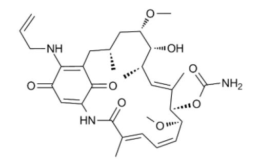
17-AAG27027
CAS Number75747-14-7
Estimated Purity≥99%
Molecular Weight585.7 Da
- SizePrice
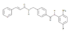
Product group Chemicals
PP24227063
CAS Number1092351-67-1
Estimated Purity≥99%
Molecular Weight308.3 Da
- SizePrice
Product group Chemicals
Chidamide27202
CAS Number743420-02-2
Estimated Purity≥98%
Molecular Weight390.4 Da
- SizePrice
Didn't find what you were looking for?
Search through our product groups to find the right product
Back to overview