

Search results: anti-Asc
Product group Antibodies

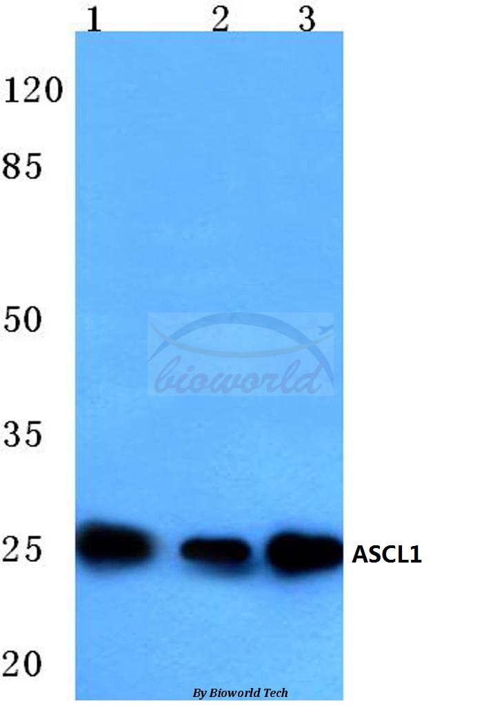
Goat anti-ASCL1 (aa79-91)EB12072
ApplicationsImmunoFluorescence, Western Blot, ELISA
TargetASCL1
- SizePrice
Product group Antibodies
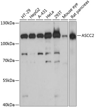
Anti-ASCC2 Antibody144-60482
ApplicationsWestern Blot, ImmunoHistoChemistry
TargetASCC2
- SizePrice
Product group Antibodies
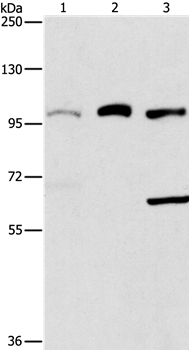
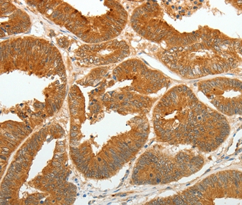
Anti-ASCC2 AntibodyA87883
ApplicationsWestern Blot, ImmunoHistoChemistry
- SizePrice
Product group Antibodies
Anti-ASCC3 AntibodyA10007
ApplicationsImmunoFluorescence, ImmunoCytoChemistry
TargetASCC3
- SizePrice

Product group Antibodies
Anti-ASCC2 AntibodyA12860
ApplicationsWestern Blot, ImmunoHistoChemistry
TargetASCC2
- SizePrice
Product group Antibodies
Anti-ASCL1 AntibodyA95934
ApplicationsWestern Blot, ELISA, ImmunoHistoChemistry
- SizePrice
Product group Antibodies
ApplicationsWestern Blot, ImmunoHistoChemistry
- SizePrice
Product group Antibodies
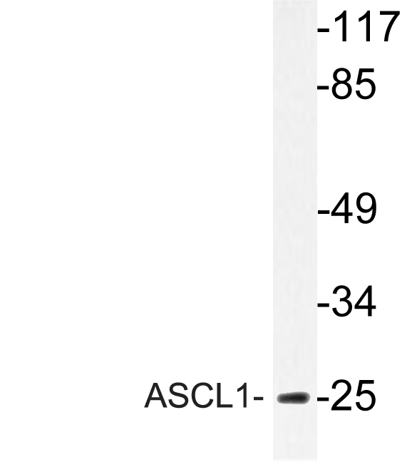
Anti-ASCL1 (E158) AntibodyA03023-1
ApplicationsWestern Blot
TargetASCL1
- SizePrice

Product group Antibodies
Anti-ASCL1 Antibody Picoband(r)A03023-2-CARRIER-FREE
ApplicationsFlow Cytometry, Western Blot, ImmunoHistoChemistry
TargetASCL1
- SizePrice
Product group Antibodies
Anti-ASCL1 Antibody Picoband(r)A03023-2-CY3
ApplicationsFlow Cytometry, Western Blot, ImmunoHistoChemistry
TargetASCL1
- SizePrice
Didn't find what you were looking for?
Search through our product groups to find the right product
Back to overview