

Search results: il-2

Product group Antibodies
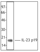
Anti-IL23/IL23A Antibody Picoband(r)A01097-1-BIOTIN
ApplicationsWestern Blot, ELISA
ReactivityHuman, Mouse, Rat
TargetIL23A
- SizePrice
Product group Antibodies
Anti-IL23/IL23A Antibody Picoband(r)A01097-1-CARRIER-FREE
ApplicationsWestern Blot, ELISA
ReactivityHuman, Mouse, Rat
TargetIL23A
- SizePrice
Product group Antibodies
Anti-IL23/IL23A Antibody Picoband(r)A01097-1-CY3
ApplicationsWestern Blot, ELISA
ReactivityHuman, Mouse, Rat
TargetIL23A
- SizePrice
Product group Antibodies
Anti-IL23/IL23A Antibody Picoband(r)A01097-1-DYLIGHT488
ApplicationsWestern Blot, ELISA
ReactivityHuman, Mouse, Rat
TargetIL23A
- SizePrice
Product group Antibodies
Anti-IL23/IL23A Antibody Picoband(r)A01097-1-DYLIGHT550
ApplicationsWestern Blot, ELISA
ReactivityHuman, Mouse, Rat
TargetIL23A
- SizePrice
Product group Antibodies
Anti-IL23/IL23A Antibody Picoband(r)A01097-1-DYLIGHT594
ApplicationsWestern Blot, ELISA
ReactivityHuman, Mouse, Rat
TargetIL23A
- SizePrice
Product group Antibodies
Anti-IL23/IL23A Antibody Picoband(r)A01097-1-FITC
ApplicationsWestern Blot, ELISA
ReactivityHuman, Mouse, Rat
TargetIL23A
- SizePrice
Product group Antibodies
Anti-IL23/IL23A Antibody Picoband(r)A01097-1-HRP
ApplicationsWestern Blot, ELISA
ReactivityHuman, Mouse, Rat
TargetIL23A
- SizePrice
Product group Antibodies
Anti-IL23/IL23A Antibody Picoband(r)A01097-1-IFLUOR647
ApplicationsWestern Blot, ELISA
ReactivityHuman, Mouse, Rat
TargetIL23A
- SizePrice
Product group Antibodies
Anti-IL23/IL23A Antibody Picoband(r)A01097-1-PE
ApplicationsWestern Blot, ELISA
ReactivityHuman, Mouse, Rat
TargetIL23A
- SizePrice
Product group Antibodies
References
ApplicationsWestern Blot, ELISA
ReactivityHuman, Mouse, Rat
TargetIL23A
- SizePrice
Product group Antibodies
ApplicationsWestern Blot, ImmunoHistoChemistry
ReactivityHuman
TargetIL23A
- SizePrice
Didn't find what you were looking for?
Search through our product groups to find the right product
Back to overview