Bio-Connect

Search results: p53 TP53

145 to 156 of 16.078 products
Sort by
Figure 1. Western blot analysis of extract from HT-29 cells untreated or treated with UV (20 min), using p53 antibody (Lane 1 and 2) and p53 pSer33 antibody (Lane 3 and 4).
Product group Antibodies
OriGene
ApplicationsImmunoFluorescence, Western Blot
ReactivityHuman
TargetTP53
  • SizePrice
Figure 1. Western blot analysis of extract from HT-29 cells untreated or treated with UV (20 min), using p53 antibody (Lane 1 and 2) and p53 pSer33 antibody (Lane 3 and 4).
Product group Antibodies
OriGene
ApplicationsImmunoFluorescence, Western Blot
ReactivityHuman
TargetTP53
  • SizePrice
Figure 1. Western blot analysis of extract from HT-29 cells untreated or treated with Doxorubicin (1mM, 30min), using p53 antibody (Lane 1 and 2) and p53 pSer37 antibody (Lane 3 and 4).
Product group Antibodies
OriGene
ApplicationsImmunoFluorescence, Western Blot
ReactivityHuman
TargetTP53
  • SizePrice
Western blot (WB) analysis of p-P53 antibody (Cat.-No.: AP20897PU-N) in extracts from COS7 cells.
Product group Antibodies
OriGene
ApplicationsWestern Blot, ImmunoHistoChemistry
ReactivityHuman, Mouse
TargetTP53
  • SizePrice
Immunohistochemistry (IHC) analyzes of p-P53 (pSer37) pAb in paraffin-embedded human lung carcinoma tissue.
Product group Antibodies
OriGene
ApplicationsImmunoHistoChemistry
ReactivityHuman
TargetTP53
  • SizePrice
Immunohistochemical analysis of paraffin-embedded human breast carcinoma tissue using p53 (Phospho-Thr387) antibody (left)or the same antibody preincubated with blocking peptide (right).
Product group Antibodies
OriGene
ApplicationsImmunoHistoChemistry
ReactivityHuman
TargetTP53
  • SizePrice
Immunohistochemical analysis of paraffin-embedded human breast carcinoma tissue using p53 (Phospho-Thr387) Antibody (left)or the same antibody preincubated with blocking peptide (right).
Product group Antibodies
OriGene
ApplicationsImmunoHistoChemistry
ReactivityHuman
TargetTP53
  • SizePrice
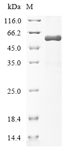
Coomassie blue staining of purified TP53 protein (Cat# TP325604). The protein was produced from HEK293T cells transfected with TP53 cDNA clone (Cat# RC225604) using MegaTran 2.0 (Cat# TT210002).
Product group Proteins / Signaling Molecules
OriGene
Protein IDK7PPA8
  • SizePrice
P53 binds to the predicted binding site (BS) for miR-138 in vitro. Electrophoretic mobility shift assay (EMSA) was performed with 100 ng p53 protein (OriGene TP300003) and biotin-labeled oligonucleotides. The p53 antibody and unlabeled competitor probe were added as indicated. Figure cited from Sci Rep, PMID: 27183959
Product group Proteins / Signaling Molecules
OriGene
Protein IDK7PPA8
  • SizePrice
Coomassie blue staining of purified TP53 protein (Cat# TP325604). The protein was produced from HEK293T cells transfected with TP53 cDNA clone (Cat# RC225604) using MegaTran 2.0 (Cat# TT210002).
Product group Proteins / Signaling Molecules
OriGene
Protein IDK7PPA8
  • SizePrice
Coomassie blue staining of purified TP53 protein (Cat# TP325604). The protein was produced from HEK293T cells transfected with TP53 cDNA clone (Cat# RC225604) using MegaTran 2.0 (Cat# TT210002).
Product group Proteins / Signaling Molecules
OriGene
Protein IDK7PPA8
  • SizePrice