Bio-Connect

Secondary

6757 to 6768 of 14.725 products
Sort by
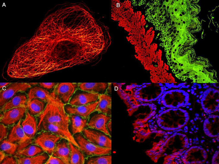
ATTO ® dyes can be used for multicolor immunofluorescent detection with low background and high signal. Examples shown are: A. Tubulin in PtK2- male Rat Kangaroo Kidney Epithelial Cells was detected using ATTO 532 labeled secondary antibody. B. Muscle alpha-actin was stained with a mouse primary antibody and ATTO 488 anti-mouse IgG (green) while Cytokeratin was stained with polyclonal rabbit anti-cytokeratin and ATTO 647N anti-rabbit IgG (red). C. HUVEC (Human umbilical vein endothelial cells were stained with anti- Vimentin-ATTO 532 (green), anti-E-Cadherin-ATTO 655 (red) and DAPI (blue). D. Rat colon sections were stained with Anti-Aquaporin 3-ATTO 594 antibody. Hoechst 33342 (blue) is used as counterstain. Images provided courtesy of Dr. Joerg Reichwein, ATTO-TEC GmbH
Product group Antibodies
OriGene
ApplicationsImmunoFluorescence, Western Blot
  • SizePrice
Western Blot of Anti-Rat IgG (H&L) (GOAT) Antibody (Min X Human Serum Proteins) (p/n 612-1125). Lane M: 3 microl Molecular Ladder. Lane 1: Rat IgG whole molecule (p/n 012-0102). Lane 2: Rat IgG F(c) Fragment (p/n 012-0103). Lane 3: Rat IgG Fab Fragment (p/n 012-0105). Lane 4: Rat IgM Whole Molecule (p/n 012-0107). Lane 5: Rat Serum (p/n D310-05). All samples were reduced. Load: 50 ng per lane. Block: MB-070 for 30 min at RT. Primary Antibody: Anti-Rat IgG (H&L) (GOAT) Antibody (Min X Human Serum Proteins) (p/n 612-1125) 1:1,000 for 60 min at RT. Secondary Antibody: Anti-Goat IgG (DONKEY) Peroxidase Conjugated Antibody (p/n CUST10) 1:40,000 in MB-070 for 30 min at RT. Predicted/Obsevered Size: 25 and 55 kDa for Rat IgG and Serum, 25 kDa for F(c) and Fab, 78 and 25 kDa for IgM. Rat F(c) migrates slightly higher.
Product group Antibodies
OriGene
ApplicationsWestern Blot, ELISA, ImmunoHistoChemistry
  • SizePrice
ATTO ® dyes can be used for multicolor immunofluorescent detection with low background and high signal. Examples shown are: A. Tubulin in PtK2- male Rat Kangaroo Kidney Epithelial Cells was detected using ATTO 532 labeled secondary antibody. B. Muscle alpha-actin was stained with a mouse primary antibody and ATTO 488 anti-mouse IgG (green) while Cytokeratin was stained with polyclonal rabbit anti-cytokeratin and ATTO 647N anti-rabbit IgG (red). C. HUVEC (Human umbilical vein endothelial cells were stained with anti- Vimentin-ATTO 532 (green), anti-E-Cadherin-ATTO 655 (red) and DAPI (blue). D. Rat colon sections were stained with Anti-Aquaporin 3-ATTO 594 antibody. Hoechst 33342 (blue) is used as counterstain. Images provided courtesy of Dr. Joerg Reichwein, ATTO-TEC GmbH
Product group Antibodies
OriGene
ApplicationsImmunoFluorescence, Western Blot
  • SizePrice

Didn't find what you were looking for?

Search through our product groups to find the right product

Back to overview
AntibodiesChemicalsProteins / Signaling MoleculesAssaysDNA / RNA / VectorsCell CultureGenome Editing and EngineeringMolecular BiologyLysates / ExtractsExpressionLabware / AccesoriesBody ProductsTechnique Specific ProductsArrays / SlidesDiagnosticBiologicalsMicrobiologyPromotions