

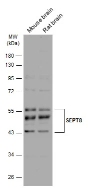
Various tissue extracts (10 μg) were separated by 10% SDS-PAGE, and the membrane was blotted with SEPT8 antibody (GTX119118) diluted at 1:4000. The HRP-conjugated anti-rabbit IgG antibody (GTX213110-01) was used to detect the primary antibody.
 dilution: 1:500.
Antigen Retrieval: Trilogy? (EDTA based, pH 8.0) buffer, 15min")
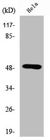
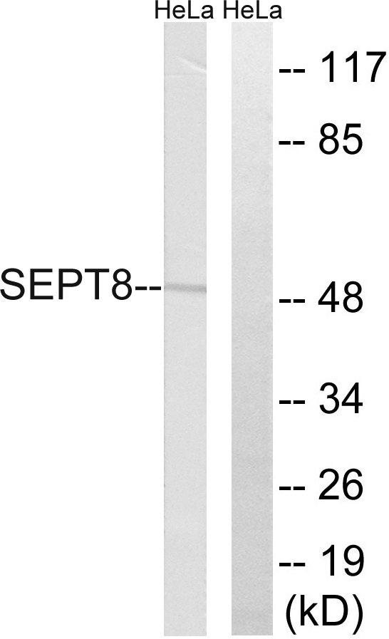
 A: HCT116 12% SDS PAGE GTX119118 diluted at 1:2000")
![SEPT8 antibody detects SEPT8 protein by immunofluorescent analysis. Sample: DIV9 rat E18 primary cortical neurons were fixed in 4% paraformaldehyde at RT for 15 min. Green: SEPT8 protein stained by SEPT8 antibody (GTX119118) diluted at 1:500. Red: beta Tubulin 3/ Tuj1, stained by beta Tubulin 3/ Tuj1 antibody [GT886] (GTX631830) diluted at 1:500. Blue: Fluoroshield with DAPI (GTX30920).](https://www.genetex.com/upload/website/prouct_img/normal/GTX119118/GTX119118_40331_20170705_IFA_R_w_23060519_148.webp "SEPT8 antibody detects SEPT8 protein by immunofluorescent analysis. Sample: DIV9 rat E18 primary cortical neurons were fixed in 4% paraformaldehyde at RT for 15 min. Green: SEPT8 protein stained by SEPT8 antibody (GTX119118) diluted at 1:500. Red: beta Tubulin 3/ Tuj1, stained by beta Tubulin 3/ Tuj1 antibody [GT886] (GTX631830) diluted at 1:500. Blue: Fluoroshield with DAPI (GTX30920).")
 of methanol-fixed HeLa, using 39332(GTX119118) antibody (Green) at 1:200 dilution. Alpha-tubulin filaments were labeled with GTX11304 (Red) at 1:2000.")
Various tissue extracts (10 μg) were separated by 10% SDS-PAGE, and the membrane was blotted with SEPT8 antibody (GTX119118) diluted at 1:4000. The HRP-conjugated anti-rabbit IgG antibody (GTX213110-01) was used to detect the primary antibody.
SEPT8 antibody
GTX119118
ApplicationsImmunoFluorescence, Western Blot, ImmunoCytoChemistry, ImmunoHistoChemistry, ImmunoHistoChemistry Paraffin
Product group Antibodies
ReactivityHuman, Mouse, Rat
TargetSEPTIN8
Overview
- SupplierGeneTex
- Product NameSEPT8 antibody
- Delivery Days Customer9
- Application Supplier NoteWB: 1:500-1:10000. ICC/IF: 1:100-1:1000. IHC-P: 1:100-1:1000. *Optimal dilutions/concentrations should be determined by the researcher.Not tested in other applications.
- ApplicationsImmunoFluorescence, Western Blot, ImmunoCytoChemistry, ImmunoHistoChemistry, ImmunoHistoChemistry Paraffin
- CertificationResearch Use Only
- ClonalityPolyclonal
- Concentration0.83 mg/ml
- ConjugateUnconjugated
- Gene ID23176
- Target nameSEPTIN8
- Target descriptionseptin 8
- Target synonymsSEP2, SEPT8, Septin-8, septin-8
- HostRabbit
- IsotypeIgG
- Protein IDQ92599
- Protein NameSeptin-8
- Scientific DescriptionSEPT8 is a member of the highly conserved septin family. Septins are 40- to 60-kD GTPases that assemble as filamentous scaffolds. They are involved in the organization of submembranous structures, in neuronal polarity, and in vesicle trafficking (Blaser et al., 2003 [PubMed 12909369]).[supplied by OMIM]
- ReactivityHuman, Mouse, Rat
- Storage Instruction-20°C or -80°C,2°C to 8°C
- UNSPSC41116161
Datasheet
Related products
Product group Antibodies
SEPT8 AntibodyCSB-PA004070
ApplicationsWestern Blot, ELISA
ReactivityHuman, Mouse, Rat
TargetSEPTIN8
- SizePrice
Product group Antibodies
Anti-SEPT8 AntibodyA97239
ApplicationsWestern Blot, ELISA
ReactivityHuman, Mouse, Rat
- SizePrice
Product group Antibodies

SEPT8 / Septin 8 AntibodyLS-C749984
ApplicationsWestern Blot
ReactivityHuman, Mouse, Rat
TargetSEPTIN8
- SizePrice

Product group Antibodies
ApplicationsFlow Cytometry, ImmunoFluorescence, ImmunoPrecipitation, Western Blot, ImmunoCytoChemistry, ImmunoHistoChemistry
ReactivityHuman, Mouse, Rat
TargetSEPTIN8
- SizePrice

Product group Antibodies
Anti-SEPT8 AntibodyHPA036534
ApplicationsWestern Blot, ImmunoCytoChemistry
ReactivityHuman
TargetSEPTIN8
- SizePrice
Product group Antibodies
Anti-SEPT9 Antibody144-08657
ApplicationsWestern Blot
ReactivityHuman, Mouse, Rat
TargetSEPTIN8
- SizePrice
Product group Antibodies
Septin 8 Polyclonal AntibodyBS-6907R
ApplicationsImmunoFluorescence, Western Blot, ELISA, ImmunoCytoChemistry, ImmunoHistoChemistry, ImmunoHistoChemistry Frozen, ImmunoHistoChemistry Paraffin
ReactivityBovine, Human, Mouse, Rat
TargetSEPTIN8
- SizePrice