

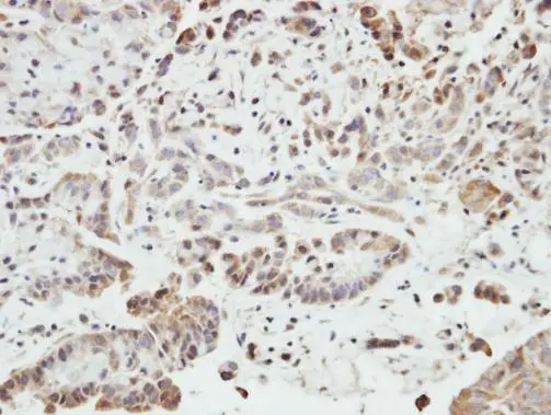
Immunohistochemical analysis of paraffin-embedded SW480 xenograft, using SHANK1(GTX107701) antibody at 1:500 dilution.
Antigen Retrieval: Trilogy? (EDTA based, pH 8.0) buffer, 15min
![SHANK1 antibody [C3], C-term detects SHANK1 protein at cell body and synaptic vesicles by immunofluorescent analysis. Sample: DIV9 rat E18 primary cortical neurons were fixed in 4% paraformaldehyde at RT for 15 min. Green: SHANK1 protein stained by SHANK1 antibody [C3], C-term (GTX107701) diluted at 1:500. Red: beta Tubulin 3/ Tuj1, a neuron cell marker, stained by beta Tubulin 3/ Tuj1 antibody [GT11710] (GTX631836) diluted at 1:500. Blue: Fluoroshield with DAPI (GTX30920).](https://www.genetex.com/upload/website/prouct_img/normal/GTX107701/GTX107701_40114_20170727_IFA_R_w_23060120_835.webp "SHANK1 antibody [C3], C-term detects SHANK1 protein at cell body and synaptic vesicles by immunofluorescent analysis. Sample: DIV9 rat E18 primary cortical neurons were fixed in 4% paraformaldehyde at RT for 15 min. Green: SHANK1 protein stained by SHANK1 antibody [C3], C-term (GTX107701) diluted at 1:500. Red: beta Tubulin 3/ Tuj1, a neuron cell marker, stained by beta Tubulin 3/ Tuj1 antibody [GT11710] (GTX631836) diluted at 1:500. Blue: Fluoroshield with DAPI (GTX30920).")
![SHANK1 antibody [C3] detects SHANK1 protein on embryonic mouse brain by immunohistochemical analysis. Sample: Frozen section of embryonic mouse brain (mE18.5). Red: SHANK1 antibody [C3] (GTX107701) diluted at 1:250. Blue: DAPI](https://www.genetex.com/upload/website/prouct_img/normal/GTX107701/GTX107701_40114_20150925_IHC-Fr_M_w_23060120_206.webp "SHANK1 antibody [C3] detects SHANK1 protein on embryonic mouse brain by immunohistochemical analysis. Sample: Frozen section of embryonic mouse brain (mE18.5). Red: SHANK1 antibody [C3] (GTX107701) diluted at 1:250. Blue: DAPI")
![SHANK1 antibody [C3], C-term detects SHANK1 protein at cytoplasm by immunofluorescent analysis. Sample: SKNSH cells were fixed in 4% paraformaldehyde at RT for 15 min. Green: SHANK1 protein stained by SHANK1 antibody [C3], C-term (GTX107701) diluted at 1:500. Blue: Hoechst 33342 staining.](https://www.genetex.com/upload/website/prouct_img/normal/GTX107701/GTX107701_40114_IFA_w_23060120_169.webp "SHANK1 antibody [C3], C-term detects SHANK1 protein at cytoplasm by immunofluorescent analysis. Sample: SKNSH cells were fixed in 4% paraformaldehyde at RT for 15 min. Green: SHANK1 protein stained by SHANK1 antibody [C3], C-term (GTX107701) diluted at 1:500. Blue: Hoechst 33342 staining.")
![SHANK1 antibody [C3], C-term detects SHANK1 protein by immunohistochemical analysis. Samples: Paraffin-Embedded mouse retina. Green: SHANK1 protein stained by SHANK1 antibody [C3], C-term (GTX107701) diluted at 1:250. Red: beta Tubulin 3/ Tuj1, stained by beta Tubulin 3/ Tuj1 antibody [GT1338] (GTX631831) diluted at 1:500. Blue: Fluoroshield with DAPI (GTX30920).
Antigen Retrieval: Citrate buffer, pH 6.0, 15 min](https://www.genetex.com/upload/website/prouct_img/normal/GTX107701/GTX107701_40114_20171017_IHC-P_M_w_23060120_895.webp "SHANK1 antibody [C3], C-term detects SHANK1 protein by immunohistochemical analysis. Samples: Paraffin-Embedded mouse retina. Green: SHANK1 protein stained by SHANK1 antibody [C3], C-term (GTX107701) diluted at 1:250. Red: beta Tubulin 3/ Tuj1, stained by beta Tubulin 3/ Tuj1 antibody [GT1338] (GTX631831) diluted at 1:500. Blue: Fluoroshield with DAPI (GTX30920).
Antigen Retrieval: Citrate buffer, pH 6.0, 15 min")
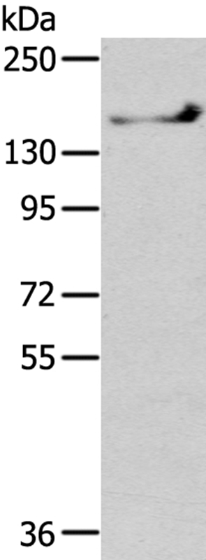
![Various whole cell extracts (30 μg) were separated by 5% SDS-PAGE, and the membrane was blotted with SHANK1 antibody [C3], C-term (GTX107701) diluted at 1:500. The HRP-conjugated anti-rabbit IgG antibody (GTX213110-01) was used to detect the primary antibody.](https://www.genetex.com/upload/website/prouct_img/normal/GTX107701/GTX107701_40114_20170901_WB_w_23060120_848.webp "Various whole cell extracts (30 μg) were separated by 5% SDS-PAGE, and the membrane was blotted with SHANK1 antibody [C3], C-term (GTX107701) diluted at 1:500. The HRP-conjugated anti-rabbit IgG antibody (GTX213110-01) was used to detect the primary antibody.")
![SHANK1 antibody [C3], C-term detects SHANK1 Protein expression by immunohistochemical analysis. Sample: Frozen-sectioned adult mouse cerebellum. Green: SHANK1 stained by SHANK1 antibody [C3], C-term (GTX107701) diluted at 1:250. Red: NF-H, stained by NF-H antibody [GT114] (GTX634289) diluted at 1:500. Blue: Fluoroshield with DAPI (GTX30920).
Antigen Retrieval: Citrate buffer, pH 6.0, 10 min](https://www.genetex.com/upload/website/prouct_img/normal/GTX107701/GTX107701_40436_20170831_IHC-Fr_M_w_23060120_480.webp "SHANK1 antibody [C3], C-term detects SHANK1 Protein expression by immunohistochemical analysis. Sample: Frozen-sectioned adult mouse cerebellum. Green: SHANK1 stained by SHANK1 antibody [C3], C-term (GTX107701) diluted at 1:250. Red: NF-H, stained by NF-H antibody [GT114] (GTX634289) diluted at 1:500. Blue: Fluoroshield with DAPI (GTX30920).
Antigen Retrieval: Citrate buffer, pH 6.0, 10 min")
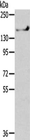
![Various whole cell extracts (30 μg) were separated by 5% SDS-PAGE, and the membrane was blotted with SHANK1 antibody [C3], C-term (GTX107701) diluted at 1:500. The HRP-conjugated anti-rabbit IgG antibody (GTX213110-01) was used to detect the primary antibody.](https://www.genetex.com/upload/website/prouct_img/normal/GTX107701/GTX107701_40114_20250411_WB_25041720_129.webp "Various whole cell extracts (30 μg) were separated by 5% SDS-PAGE, and the membrane was blotted with SHANK1 antibody [C3], C-term (GTX107701) diluted at 1:500. The HRP-conjugated anti-rabbit IgG antibody (GTX213110-01) was used to detect the primary antibody.")
![Boiled and unboiled various tissue extracts (50 μg) were separated by 5% SDS-PAGE, and the membrane was blotted with SHANK1 antibody [C3], C-term (GTX107701) diluted at 1:1000. The HRP-conjugated anti-rabbit IgG antibody (GTX213110-01) was used to detect the primary antibody.](https://www.genetex.com/upload/website/prouct_img/normal/GTX107701/GTX107701_40114_20250418_WB_M_tissue_ub_25042420_693.webp "Boiled and unboiled various tissue extracts (50 μg) were separated by 5% SDS-PAGE, and the membrane was blotted with SHANK1 antibody [C3], C-term (GTX107701) diluted at 1:1000. The HRP-conjugated anti-rabbit IgG antibody (GTX213110-01) was used to detect the primary antibody.")
Immunohistochemical analysis of paraffin-embedded SW480 xenograft, using SHANK1(GTX107701) antibody at 1:500 dilution.
Antigen Retrieval: Trilogy? (EDTA based, pH 8.0) buffer, 15min
SHANK1 antibody [C3], C-term
GTX107701
ApplicationsImmunoFluorescence, Western Blot, ImmunoCytoChemistry, ImmunoHistoChemistry, ImmunoHistoChemistry Frozen, ImmunoHistoChemistry Paraffin
Product group Antibodies
ReactivityHuman, Mouse, Rat
TargetSHANK1
Overview
- SupplierGeneTex
- Product NameSHANK1 antibody [C3], C-term
- Delivery Days Customer9
- Application Supplier NoteWB: 1:500-1:3000. ICC/IF: 1:100-1:1000. IHC-P: 1:100-1:1000. IHC-Fr: 1:100-1:1000. *Optimal dilutions/concentrations should be determined by the researcher.Not tested in other applications.
- ApplicationsImmunoFluorescence, Western Blot, ImmunoCytoChemistry, ImmunoHistoChemistry, ImmunoHistoChemistry Frozen, ImmunoHistoChemistry Paraffin
- CertificationResearch Use Only
- ClonalityPolyclonal
- Concentration0.53 mg/ml
- ConjugateUnconjugated
- Gene ID50944
- Target nameSHANK1
- Target descriptionSH3 and multiple ankyrin repeat domains 1
- Target synonymsSPANK-1, SSTRIP, synamon, SH3 and multiple ankyrin repeat domains protein 1, SSTR-interacting protein, somatostatin receptor-interacting protein
- HostRabbit
- IsotypeIgG
- Protein IDQ9Y566
- Protein NameSH3 and multiple ankyrin repeat domains protein 1
- Scientific DescriptionSeems to be an adapter protein in the postsynaptic density (PSD) of excitatory synapses that interconnects receptors of the postsynaptic membrane including NMDA-type and metabotropic glutamate receptors via complexes with GKAP/PSD-95 and Homer, respectively, and the actin-based cytoskeleton. May play a role in the structural and functional organization of the dendritic spine and synaptic junction.
- ReactivityHuman, Mouse, Rat
- Storage Instruction-20°C or -80°C,2°C to 8°C
- UNSPSC41116161
Datasheet
Related products
Product group Antibodies
Anti-SHANK1 AntibodyA42540
ApplicationsWestern Blot
ReactivityHuman, Mouse
- SizePrice
Product group Antibodies

SHANK1 AntibodyLS-C754236
ApplicationsWestern Blot, ELISA
ReactivityHuman
TargetSHANK1
- SizePrice

Product group Antibodies
Anti-SHANK1 AntibodyHPA032129
ApplicationsWestern Blot, ImmunoCytoChemistry, ImmunoHistoChemistry
ReactivityHuman
TargetSHANK1
- SizePrice
Product group Antibodies
SHANK1 AntibodyCSB-PA100838
ApplicationsWestern Blot, ELISA
ReactivityHuman, Mouse, Rat
TargetSHANK1
- SizePrice
Product group Antibodies
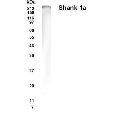
SHANK1a antibodyGTX70380
ApplicationsWestern Blot, ImmunoHistoChemistry
ReactivityHuman, Rat
TargetSHANK1
- SizePrice
Product group Antibodies
SHANK1 Polyclonal AntibodyBS-0211R
ApplicationsImmunoFluorescence, Western Blot, ELISA, ImmunoCytoChemistry, ImmunoHistoChemistry, ImmunoHistoChemistry Frozen, ImmunoHistoChemistry Paraffin
ReactivityHuman, Mouse, Rat
TargetSHANK1
- SizePrice
Product group Antibodies
SHANK1 antibodyGTX30444
ApplicationsWestern Blot
ReactivityHuman
TargetSHANK1
- SizePrice