

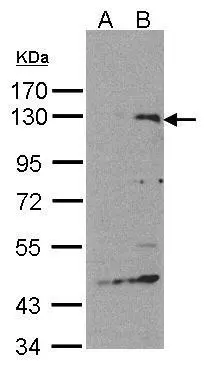
Western Blot analysis of SIPA1 expression in transfected 293T cell line by SIPA1 polyclonal antibody. A: Non-transfected lysate. B: SIPA1 transfected lysate. 7.5% SDS PAGE GTX104786 diluted at 1:500
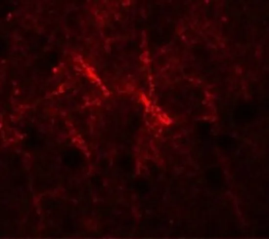
![SIPA1 antibody [C3], C-term detects SIPA1 protein at weak membrane and cytosol on Cal27 xenograft by immunohistochemical analysis. Sample: Paraffin-embedded Cal27 xenograft. SIPA1 antibody [C3], C-term (GTX104786) dilution: 1:500.
Antigen Retrieval: Trilogy? (EDTA based, pH 8.0) buffer, 15min](https://www.genetex.com/upload/website/prouct_img/normal/GTX104786/GTX104786_39680_IHC_w_23060120_361.webp "SIPA1 antibody [C3], C-term detects SIPA1 protein at weak membrane and cytosol on Cal27 xenograft by immunohistochemical analysis. Sample: Paraffin-embedded Cal27 xenograft. SIPA1 antibody [C3], C-term (GTX104786) dilution: 1:500.
Antigen Retrieval: Trilogy? (EDTA based, pH 8.0) buffer, 15min")
 antibody at 1:200 dilution.")
![SIPA1 antibody [C3], C-term detects SIPA1 protein at weak membrane and cytosol on HBL435 xenograft by immunohistochemical analysis. Sample: Paraffin-embedded HBL435 xenograft. SIPA1 antibody [C3], C-term (GTX104786) dilution: 1:500.
Antigen Retrieval: Trilogy? (EDTA based, pH 8.0) buffer, 15min](https://www.genetex.com/upload/website/prouct_img/normal/GTX104786/GTX104786_39680_IHC_2_w_23060120_932.webp "SIPA1 antibody [C3], C-term detects SIPA1 protein at weak membrane and cytosol on HBL435 xenograft by immunohistochemical analysis. Sample: Paraffin-embedded HBL435 xenograft. SIPA1 antibody [C3], C-term (GTX104786) dilution: 1:500.
Antigen Retrieval: Trilogy? (EDTA based, pH 8.0) buffer, 15min")
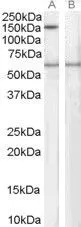
Western Blot analysis of SIPA1 expression in transfected 293T cell line by SIPA1 polyclonal antibody. A: Non-transfected lysate. B: SIPA1 transfected lysate. 7.5% SDS PAGE GTX104786 diluted at 1:500
SIPA1 antibody [C3], C-term
GTX104786
ApplicationsImmunoFluorescence, Western Blot, ImmunoCytoChemistry, ImmunoHistoChemistry, ImmunoHistoChemistry Paraffin
Product group Antibodies
ReactivityHuman, Mouse
TargetSIPA1
Overview
- SupplierGeneTex
- Product NameSIPA1 antibody [C3], C-term
- Delivery Days Customer9
- Application Supplier NoteWB: 1:500-1:3000. ICC/IF: 1:100-1:1000. IHC-P: 1:100-1:1000. *Optimal dilutions/concentrations should be determined by the researcher.Not tested in other applications.
- ApplicationsImmunoFluorescence, Western Blot, ImmunoCytoChemistry, ImmunoHistoChemistry, ImmunoHistoChemistry Paraffin
- CertificationResearch Use Only
- ClonalityPolyclonal
- Concentration1 mg/ml
- ConjugateUnconjugated
- Gene ID6494
- Target nameSIPA1
- Target descriptionsignal-induced proliferation-associated 1
- Target synonymsSPA1, signal-induced proliferation-associated protein 1, GTPase-activating protein Spa-1, p130 SPA-1, signal-induced proliferation-associated gene 1, sipa-1
- HostRabbit
- IsotypeIgG
- Protein IDQ96FS4
- Protein NameSignal-induced proliferation-associated protein 1
- Scientific DescriptionThe product of this gene is a mitogen induced GTPase activating protein (GAP). It exhibits a specific GAP activity for Ras-related regulatory proteins Rap1 and Rap2, but not for Ran or other small GTPases. This protein may also hamper mitogen-induced cell cycle progression when abnormally or prematurely expressed. It is localized to the perinuclear region. Two alternatively spliced variants encoding the same isoform have been characterized to date. [provided by RefSeq]
- ReactivityHuman, Mouse
- Storage Instruction-20°C or -80°C,2°C to 8°C
- UNSPSC41116161
Datasheet
Related products
Product group Antibodies
SIPA1 AntibodyCSB-PA021330LA01HU
ApplicationsImmunoFluorescence, Western Blot, ELISA, ImmunoHistoChemistry
ReactivityHuman
TargetSIPA1
- SizePrice
Product group Antibodies
Anti-SIPA1 Antibody144-12253
ApplicationsWestern Blot
ReactivityHuman, Mouse
TargetSIPA1
- SizePrice

Product group Antibodies
Anti-SIPA1 AntibodyA46157
ApplicationsImmunoHistoChemistry
ReactivityHuman
- SizePrice
Product group Antibodies

Goat anti-SPA1 / SIPA1EB06260
ApplicationsWestern Blot, ELISA
ReactivityHuman
TargetSIPA1
- SizePrice

Product group Antibodies
Anti-SIPA1 AntibodyHPA039867
ApplicationsImmunoCytoChemistry, ImmunoHistoChemistry
ReactivityHuman
TargetSIPA1
- SizePrice
Product group Antibodies

SIPA1 AntibodyLS-C406036
ApplicationsELISA, ImmunoHistoChemistry
ReactivityHuman
TargetSIPA1
- SizePrice
Product group Antibodies
SIPA1 Polyclonal AntibodyCAC15063
ApplicationsImmunoFluorescence, Western Blot, ELISA, ImmunoHistoChemistry
TargetSIPA1
- SizePrice
Product group Antibodies
SPA-1 Polyclonal AntibodyBS-1602R
ApplicationsImmunoFluorescence, ELISA, ImmunoCytoChemistry, ImmunoHistoChemistry, ImmunoHistoChemistry Frozen, ImmunoHistoChemistry Paraffin
ReactivityBovine, Canine, Equine, Human, Mouse, Porcine, Rat
TargetSIPA1
- SizePrice
Product group Antibodies
SIPA1 antibody, C-termGTX10162
ApplicationsWestern Blot
ReactivityHuman
TargetSIPA1
- SizePrice
Product group Antibodies
SIPA1 antibodyGTX31846
ApplicationsWestern Blot, ELISA, ImmunoHistoChemistry, ImmunoHistoChemistry Paraffin
ReactivityHuman
TargetSIPA1
- SizePrice