

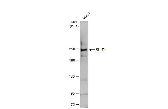
Whole cell extract (30 μg) was separated by 5% SDS-PAGE, and the membrane was blotted with SLIT1 antibody (GTX134122) diluted at 1:1000. The HRP-conjugated anti-rabbit IgG antibody (GTX213110-01) was used to detect the primary antibody.
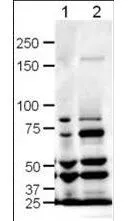
 were separated by 5% SDS-PAGE, and the membrane was blotted with SLIT1 antibody (GTX134122) diluted at 1:500. The HRP-conjugated anti-rabbit IgG antibody (GTX213110-01) was used to detect the primary antibody.")
![SLIT1 antibody detects SLIT1 protein by immunofluorescent analysis. Sample: DIV9 rat E18 primary cortical neuron cells were fixed in 4% paraformaldehyde at RT for 15 min. Green: SLIT1 stained by SLIT1 antibody (GTX134122) diluted at 1:500. Red: Tau, stained by Tau antibody [GT287] (GTX634809) diluted at 1:500. Blue: Fluoroshield with DAPI (GTX30920).](https://www.genetex.com/upload/website/prouct_img/normal/GTX134122/GTX134122_43243_20190306_ICC_IF_R_w_23060620_100.webp "SLIT1 antibody detects SLIT1 protein by immunofluorescent analysis. Sample: DIV9 rat E18 primary cortical neuron cells were fixed in 4% paraformaldehyde at RT for 15 min. Green: SLIT1 stained by SLIT1 antibody (GTX134122) diluted at 1:500. Red: Tau, stained by Tau antibody [GT287] (GTX634809) diluted at 1:500. Blue: Fluoroshield with DAPI (GTX30920).")
Whole cell extract (30 μg) was separated by 5% SDS-PAGE, and the membrane was blotted with SLIT1 antibody (GTX134122) diluted at 1:1000. The HRP-conjugated anti-rabbit IgG antibody (GTX213110-01) was used to detect the primary antibody.
SLIT1 antibody
GTX134122
ApplicationsImmunoFluorescence, Western Blot, ImmunoCytoChemistry
Product group Antibodies
ReactivityHuman, Mouse, Rat
TargetSlit1
Overview
- SupplierGeneTex
- Product NameSLIT1 antibody
- Delivery Days Customer9
- Application Supplier NoteWB: 1:500-1:3000. ICC/IF: 1:100-1:1000. *Optimal dilutions/concentrations should be determined by the researcher.Not tested in other applications.
- ApplicationsImmunoFluorescence, Western Blot, ImmunoCytoChemistry
- CertificationResearch Use Only
- ClonalityPolyclonal
- Concentration0.81 mg/ml
- ConjugateUnconjugated
- Gene ID20562
- Target nameSlit1
- Target descriptionslit guidance ligand 1
- Target synonymsSlil1, mKIAA0813, slit homolog 1 protein, slit-1
- HostRabbit
- IsotypeIgG
- Protein IDQ80TR4
- Protein NameSlit homolog 1 protein
- ReactivityHuman, Mouse, Rat
- Storage Instruction-20°C or -80°C,2°C to 8°C
- UNSPSC41116161
Datasheet
Related products
Product group Antibodies
ApplicationsImmunoPrecipitation, Western Blot, ImmunoCytoChemistry, ImmunoHistoChemistry
ReactivityMouse, Rat
TargetSlit1
- SizePrice
Product group Antibodies
SLIT1 antibodyGTX48742
ApplicationsWestern Blot, ELISA, ImmunoHistoChemistry, ImmunoHistoChemistry Paraffin
ReactivityMouse, Rat
TargetSlit1
- SizePrice