

Snx6 Polyclonal Antibody
CAC09931
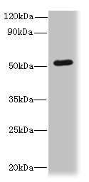
ApplicationsWestern Blot, ELISA, ImmunoHistoChemistry
Product group Antibodies
TargetSNX6
Overview
- SupplierBiomatik
- Product NameSnx6 Polyclonal Antibody
- Delivery Days Customer12
- ApplicationsWestern Blot, ELISA, ImmunoHistoChemistry
- Applications SupplierELISA, WB, IHC; Recommended dilution: WB:1:500-1:2000, IHC:1:20-1:200
- CertificationResearch Use Only
- ClonalityPolyclonal
- ConjugateUnconjugated
- Gene ID58533
- Target nameSNX6
- Target descriptionsorting nexin 6
- Target synonymsMSTP010, TFAF2, sorting nexin-6, TRAF4-associated factor 2, tumor necrosis factor receptor-associated factor 4(TRAF4)-associated factor 2
- HostRabbit
- IsotypeIgG
- Protein IDQ9UNH7
- Protein NameSorting nexin-6
- Scientific DescriptionThe Snx6 Polyclonal Antibody (Species: Human) has been validated for the following applications: ELISA, WB, IHC.
- Reactivity SupplierHuman
- Storage Instruction-20°C,2°C to 8°C
- UNSPSC12352203
Related products

Product group Antibodies
Anti-SNX6 Antibody Picoband(r)A06545-CARRIER-FREE
ApplicationsFlow Cytometry, ImmunoFluorescence, Western Blot, ELISA, ImmunoCytoChemistry
ReactivityHuman, Mouse, Rat
TargetSNX6
- SizePrice
Product group Antibodies
References
SNX6 Polyclonal AntibodyBS-12410R
ApplicationsImmunoFluorescence, Western Blot, ELISA, ImmunoCytoChemistry, ImmunoHistoChemistry, ImmunoHistoChemistry Frozen, ImmunoHistoChemistry Paraffin
ReactivityBovine, Chicken, Equine, Human, Mouse, Porcine, Rabbit, Rat, Sheep
TargetSNX6
- SizePrice

Product group Antibodies
Anti-SNX6 AntibodyHPA049374
ApplicationsWestern Blot, ImmunoCytoChemistry, ImmunoHistoChemistry
ReactivityHuman
TargetSNX6
- SizePrice
Product group Antibodies
SNX6 AntibodyCSB-PA890766LA01HU
ApplicationsWestern Blot, ELISA, ImmunoHistoChemistry
ReactivityHuman
TargetSNX6
- SizePrice
Product group Antibodies

SNX6 AntibodyLS-C830010
ApplicationsELISA, ImmunoHistoChemistry
ReactivityHuman, Mouse
TargetSNX6
- SizePrice